Spinal Muscular Atrophy (SMA)Spinal Cord-Related Muscle Wasting (Spinal Muscular Atrophy (SMA))
Spinal Muscular Atrophy (SMA) — Overview
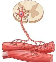
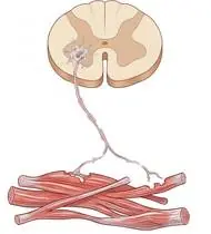
Spinal Muscular Atrophy (SMA) is an autosomal recessive neuromuscular disorder caused by homozygous deletion/mutation of the SMN1 (Survival Motor Neuron 1) gene on chromosome 5q13 (with phenotypic severity modified by SMN2 copy number), leading to progressive degeneration of spinal cord anterior horn alpha motor neurons. It produces symmetric, proximal > distal lower motor neuron weakness (lower extremities affected more than upper extremities), hypotonia, areflexia, intercostal muscle weakness (with relative diaphragmatic sparing!), and neuromuscular scoliosis, while cognition and sensation are 100% normal. In the modern era of disease-modifying therapies (Nusinersen, Onasemnogene abeparvovec, Risdiplam), proactive physiotherapy maximizes motor function!
Spinal Muscular Atrophy (SMA) is a genetic motor nerve condition in which a lack of the SMN protein weakens the motor nerve cells in the spinal cord—affecting the proximal muscles closest to the trunk (hips, thighs, shoulders, and back) while leaving intelligence, emotional warmth, and touch sensation completely bright and intact. Especially alongside modern SMA treatments, daily stretching, posture care, and gentle non-fatiguing exercise help unlock and preserve every motor skill!
Primary Causes & Biomechanics
Key Points You Should Know
- •SMN protein deficiency causing selective loss of spinal alpha motor neurons (pure Lower Motor Neuron syndrome—zero spasticity!)
- •Clinical classification by highest motor milestone: Type 1 (Werdnig-Hoffmann: non-sitters, <6 mos), Type 2 (sitters, 6–18 mos), Type 3 (Kugelberg-Welander: walkers, >18 mos), and Type 4 (adult-onset)
- •Intercostal Weakness vs. Diaphragm Sparing: Weak intercostal muscles combined with a strong diaphragm create paradoxical breathing, pectus excavatum/bell-shaped chest, and impaired cough clearance
- •Why Hip & Knee Stretching + Standing Frames Matter So Much: Sitting for long periods allows the hips, knees, and ankles to tighten into bent contractures and allows the spine to curve. Daily gentle stretching, night splints, and supported standing frames keep the joints straight and bones strong.
- •Protecting the Ribcage & Cough: In SMA, the diaphragm muscle at the bottom of the lungs is stronger than the rib muscles between the ribs—practicing deep rib-expanding breaths helps keep the chest wall wide and clear.
- •Water Is Your Superpower (Hydrotherapy): In a warm pool, buoyancy lifts the weight of gravity off your legs and arms so you can move freely without tiring out.
Assessment Methods
1. Goniometric ROM & Postural / Kinematic Analysis
HFMSE (Hammersmith Functional Motor Scale Expanded) for Types 2/3, CHOP INTEND for Type 1, RULM (Revised Upper Limb Module), and joint contracture goniometry (hip/knee flexion and ankle equinus).
2. Special Clinical Provocation & Neurological/Functional Tests
6-Minute Walk Test (6MWT — monitoring fatigability between minute 1 and minute 6!), Forced Vital Capacity (FVC), Peak Cough Flow, and Cobb angle scoliosis screening.
3. Standardized Clinical Outcome Scales & Imaging Criteria
HFMSE, RULM, CHOP INTEND, 6MWT, and FVC / Peak Cough Flow.
1. Range of Motion & Posture Check
Your physiotherapist gently evaluates how comfortably and smoothly your spinal cord anterior horn alpha motor neurons and brainstem motor nuclei (SMN1 gene deletion on chromosome 5q13) moves and checks your overall posture.
2. Muscle Strength & Functional Movement Check
Your muscle balance, walking or reaching comfort, and specific gentle movement tests are evaluated without triggering sharp pain.
3. Typical Symptoms & Daily Impact Review
Symmetric Proximal Muscle Weakness (Legs > Arms): Difficulty rising from the floor or a low chair, climbing stairs, running, or lifting arms overhead, accompanied by decreased muscle tone (hypotonia) and absent tendon reflexes.
Treatment Options & Phase-Based Protocol
✨ Enriched GIF Exercise & Precaution GuidePhase 1: Acute Symptom Modulation, Tissue Protection & Early Activation
- •Phase 1 Clinical Focus & Tissue Protection: Contracture Prevention & Respiratory Expansion Phase: Daily gentle passive/active-assisted gastro-soleus towel stretching, hip flexor/hamstring lengthening (prone positioning or resting orthoses), and thoracic expansion & diaphragmatic/costal breathing (plus breath-stacking / Cough Assist).
- •Pain, Edema & Neuromuscular Modulation: Targeted physical modalities, relative load management, and early protected mobility of the spinal cord anterior horn alpha motor neurons and brainstem motor nuclei (SMN1 gene deletion on chromosome 5q13) without provoking mechanical or inflammatory irritation.
- •Proximal & Distal Kinetic Chain Maintenance: Maintain active mobility and circulation in adjacent joints while protecting the primary healing structures in Spinal Muscular Atrophy (SMA).
Phase 2: Progressive Mobility, Motor Control & Targeted Strengthening
- •Phase 2 Progressive Mobilization & Motor Control: Submaximal Proximal & Trunk Stabilization Phase: Non-fatiguing supine heel slides, pelvic bridging, wand-assisted / active-assisted upper limb elevation, and thumb/hand dexterity (keeping effort at light-to-moderate Borg RPE 11–12).
- •Biomechanical Alignment & Co-Contraction: Restore balanced agonist-antagonist force couples, joint centration, and multi-planar neuromuscular control across the spinal cord anterior horn alpha motor neurons and brainstem motor nuclei (SMN1 gene deletion on chromosome 5q13).
- •Myofascial Extensibility & Endurance: Address secondary compensatory tightness and build local muscular endurance within safe, symptom-free biomechanical parameters.
Phase 3: Functional Integration, Proprioception & Long-Term Prevention
- •Phase 3 Functional Integration & Long-Term Prevention: Functional Transfer, Standing Frame & Ambulation Phase: Supported standing frame use (5 days/week for bone density, acetabular development, and contracture prevention in non-walkers!), side-lying/standing hip abduction, chair sit-to-stand, and aquatic therapy (hydrotherapy buoyancy allows gravity-free movement!).
- •Proprioceptive Balance & Dynamic Loading: Progress to task-specific closed-chain, balance, and functional movement patterns tailored to daily life, occupational, or athletic demands.
- •Self-Management & Relapse Prophylaxis: Establish lifelong ergonomic habits, home exercise maintenance, and early warning-sign awareness to prevent recurrence.
Step 1: Calming Pain, Protecting the Area & Gentle Starter Motion
- •Why Hip & Knee Stretching + Standing Frames Matter So Much: Sitting for long periods allows the hips, knees, and ankles to tighten into bent contractures and allows the spine to curve. Daily gentle stretching, night splints, and supported standing frames keep the joints straight and bones strong.
- •Calm Protection & Gentle Start: During the initial stage of Spinal Cord-Related Muscle Wasting, avoid painful overloading and perform the gentle, pain-free starter movements shown below to support circulation and comfort.
- •Respecting Your Body's Signals: Every movement should stay within a comfortable, non-painful zone—never force through sharp or radiating pain.
Step 2: Restoring Flexibility & Building Supporting Muscle Strength
- •Protecting the Ribcage & Cough: In SMA, the diaphragm muscle at the bottom of the lungs is stronger than the rib muscles between the ribs—practicing deep rib-expanding breaths helps keep the chest wall wide and clear.
- •Step-by-Step Flexibility & Support: As acute sensitivity settles, gradually build mobility and strengthen the supporting muscles around the joint at a steady, controlled pace.
- •Posture & Breathing Coordination: Keep your posture upright and breathe smoothly (never hold your breath!) during every repetition.
Step 3: Safe Daily Habits, Stamina & Preventing Recurrence
- •Water Is Your Superpower (Hydrotherapy): In a warm pool, buoyancy lifts the weight of gravity off your legs and arms so you can move freely without tiring out.
- •Confident Return to Daily Life: Integrate your stronger muscles and improved balance into walking, stairs, work, and recreational activities.
- •Preventing Future Flare-Ups: Continue your core protective habits and key maintenance exercises even after symptoms resolve.
Animated (GIF) Therapeutic Exercise Guide
Animated clinical exercise protocol with biomechanical angles, target muscle groups, and dosage parameters (Click image to enlarge)
Step-by-step animated exercise demonstrations you can safely perform at home (Click image to enlarge)
 🔍 Enlarge
1. Supine Active-Assisted Heel Slide & Synovial Diffusion Mobilization
1. Supine Heel Slide (Gentle Knee & Hip Bending)
Controlled flexion and extension of the lower extremity by sliding the heel along the support surface without axial weight-bearing. Stimulates synovial fluid diffusion across avascular cartilage and prevents capsular adhesions. Specifically indicated in Spinal Muscular Atrophy (SMA) rehabilitation to optimize spinal cord anterior horn alpha motor neurons and brainstem motor nuclei (SMN1 gene deletion on chromosome 5q13) biomechanics and neuromuscular control.
While lying on your back, slowly slide your heel along the bed sheet toward your hip within a comfortable, pain-free angle, hold for 3 seconds, and gently slide it back out straight. Perform smoothly within a comfortable, pain-free range to support your Spinal Cord-Related Muscle Wasting recovery.
 🔍 Enlarge
2. Long-Sitting Towel-Assisted Gastrocnemius-Soleus & Plantar Fascia Stretch
2. Seated Towel Calf & Foot Arch Stretch
Long-sitting with a towel looped around the metatarsal heads, applying a slow, sustained passive pull into ankle dorsiflexion. Restores gastrocnemius-soleus-Achilles length and reduces plantar fascial and equine tension. Specifically indicated in Spinal Muscular Atrophy (SMA) rehabilitation to optimize spinal cord anterior horn alpha motor neurons and brainstem motor nuclei (SMN1 gene deletion on chromosome 5q13) biomechanics and neuromuscular control.
Sit with your leg extended and loop a towel around the ball of your foot. Gently pull both ends of the towel toward you until you feel a comfortable stretch in your calf and sole, holding for 20–30 seconds. Perform smoothly within a comfortable, pain-free range to support your Spinal Cord-Related Muscle Wasting recovery.
 🔍 Enlarge
3. Thoracic Expansion Exercise (TEE) with Bilateral Arm Elevation & 3s Inspiratory Hold
3. Chest-Opening Deep Breath with Arm Raise
Synchronizing deep nasal inspiration with bilateral shoulder flexion/abduction and a 3-second end-inspiratory hold before passive/pursed-lip expiration. Opens collateral ventilation channels to prevent basal atelectasis and maintains costovertebral compliance. Specifically indicated in Spinal Muscular Atrophy (SMA) rehabilitation to optimize spinal cord anterior horn alpha motor neurons and brainstem motor nuclei (SMN1 gene deletion on chromosome 5q13) biomechanics and neuromuscular control.
While sitting upright, breathe in deeply through your nose while opening your arms upward and outward to expand your ribcage. Hold the breath inside for 3 seconds, then exhale slowly while lowering your arms. Perform smoothly within a comfortable, pain-free range to support your Spinal Cord-Related Muscle Wasting recovery.
 🔍 Enlarge
4. Scapulothoracic Retraction & Depression (Middle/Lower Trapezius & Rhomboids)
4. Shoulder Blade Squeeze (Postural Alignment)
Adduction and depression of the scapulae via the middle/lower trapezius and rhomboids while inhibiting upper trapezius overactivity. Corrects anterior scapular tilt, widens the subacromial outlet, and unloads the anterior thoracic column. Specifically indicated in Spinal Muscular Atrophy (SMA) rehabilitation to optimize spinal cord anterior horn alpha motor neurons and brainstem motor nuclei (SMN1 gene deletion on chromosome 5q13) biomechanics and neuromuscular control.
Sit or stand tall. Keep your shoulders relaxed away from your ears, gently draw your shoulder blades together and slightly downward toward your back pockets, hold for 5 seconds, and release. Perform smoothly within a comfortable, pain-free range to support your Spinal Cord-Related Muscle Wasting recovery.
 🔍 Enlarge
5. Supine Wand-Assisted (Cane) Active-Assisted Shoulder Elevation
5. Lying Cane/Stick-Assisted Two-Handed Arm Raise
In supine (scapula stabilized against the plinth and gravity eliminated in the final arc), the unaffected upper extremity guides a wand/stick to elevate the affected shoulder smoothly without deltoid-induced superior humeral shear. Specifically indicated in Spinal Muscular Atrophy (SMA) rehabilitation to optimize spinal cord anterior horn alpha motor neurons and brainstem motor nuclei (SMN1 gene deletion on chromosome 5q13) biomechanics and neuromuscular control.
Lie on your back holding a light stick or cane with both hands. Use your healthy arm to gently guide both arms up overhead as far as comfortable, hold for 3 seconds, and slowly lower. Perform smoothly within a comfortable, pain-free range to support your Spinal Cord-Related Muscle Wasting recovery.
 🔍 Enlarge
6. Diaphragmatic & Pursed-Lip Breathing (PLB — Autonomic & Ventilatory Control)
6. Calm Belly Breathing with Pursed-Lip Exhale
Slow nasal inhalation expanding the lower ribcage and abdomen (while relaxing accessory neck/shoulder muscles), followed by prolonged expiration through pursed lips at a 1:2 ratio. Prevents dynamic airway collapse, improves basal ventilation, and reduces sympathetic tone. Specifically indicated in Spinal Muscular Atrophy (SMA) rehabilitation to optimize spinal cord anterior horn alpha motor neurons and brainstem motor nuclei (SMN1 gene deletion on chromosome 5q13) biomechanics and neuromuscular control.
Sit comfortably and place one hand on your chest and the other on your belly. Breathe in slowly through your nose for 2–3 seconds so your belly gently rises, then breathe out slowly for 4–6 seconds through pursed lips as if flickering a candle. Perform smoothly within a comfortable, pain-free range to support your Spinal Cord-Related Muscle Wasting recovery.
Movements to Avoid & Strict Precautions
1. Exhausting High-Resistance or Eccentric Exercises (Causes Motor Unit Fatigue!)
1. Exercising to the point of muscle trembling or exhaustion (always use light, comfortable reps with plenty of rest)
2. Prolonged Sitting All Day Without Daily Prone Lying / Stretching or Night Splints
2. Sitting all day without stretching the hips, knees, and ankles straight
3. Slouched Asymmetric Sitting Without Thoracic/Scoliosis Postural Support
3. Sitting leaned over to one side without proper seating or brace support
4. Rough or Forceful Twisting Transfers (Osteopenic Bone Fracture Risk!)
4. Lifting or transferring without supporting the thighs and trunk smoothly
Spinal Muscular Atrophy (SMA) — Video Guide
Click directly on any video to open and watch in a window, or review details via 📋 Summary & Options.
Condition Overview Video (1 Video)
Summary of pathophysiology, biomechanics, and clinical presentation
What the condition is, why it develops, and its basic mechanism

What Is Spinal Muscular Atrophy (SMA)? Types and Clinical Features
Spinal Muscular Atrophy (SMA) is an autosomal recessive neuromuscular disorder caused by homozygous deletion/mutation of the SMN1 (Survival Motor Neuron 1) gene on chromosome 5q13 (with phenotypic severity modified by SMN2 copy number), leading to progressive degeneration of spinal cord anterior horn alpha motor neurons. It produces symmetric, proximal > distal lower motor neuron weakness (lower extremities affected more than upper extremities), hypotonia, areflexia, intercostal muscle weakness (with relative diaphragmatic sparing!), and neuromuscular scoliosis, while cognition and sensation are 100% normal. In the modern era of disease-modifying therapies (Nusinersen, Onasemnogene abeparvovec, Risdiplam), proactive physiotherapy maximizes motor function!
Spinal Muscular Atrophy (SMA) is a genetic motor nerve condition in which a lack of the SMN protein weakens the motor nerve cells in the spinal cord—affecting the proximal muscles closest to the trunk (hips, thighs, shoulders, and back) while leaving intelligence, emotional warmth, and touch sensation completely bright and intact. Especially alongside modern SMA treatments, daily stretching, posture care, and gentle non-fatiguing exercise help unlock and preserve every motor skill!
Treatment and Exercise Videos
Phase-based physiotherapy protocols, mobilization, and therapeutic exercises
Step-by-step recovery and safe home exercise applications

Positioning, Contracture Prevention, and Movement Training in SMA Rehab (Part 1)
Phase 1: Acute Symptom Modulation, Tissue Protection & Early Activation: Phase 1 Clinical Focus & Tissue Protection: Contracture Prevention & Respiratory Expansion Phase: Daily gentle passive/active-assisted gastro-soleus towel stretching, hip flexor/hamstring lengthening (prone positioning or resting orthoses), and thoracic expansion & diaphragmatic/costal breathing (plus breath-stacking / Cough Assist).
Step 1: Calming Pain, Protecting the Area & Gentle Starter Motion: Why Hip & Knee Stretching + Standing Frames Matter So Much: Sitting for long periods allows the hips, knees, and ankles to tighten into bent contractures and allows the spine to curve. Daily gentle stretching, night splints, and supported standing frames keep the joints straight and bones strong.

Trunk Control, Respiratory Support, and Functional Exercises in SMA (Part 6)
Phase 2: Progressive Mobility, Motor Control & Targeted Strengthening: Phase 2 Progressive Mobilization & Motor Control: Submaximal Proximal & Trunk Stabilization Phase: Non-fatiguing supine heel slides, pelvic bridging, wand-assisted / active-assisted upper limb elevation, and thumb/hand dexterity (keeping effort at light-to-moderate Borg RPE 11–12).
Step 2: Restoring Flexibility & Building Supporting Muscle Strength: Protecting the Ribcage & Cough: In SMA, the diaphragm muscle at the bottom of the lungs is stronger than the rib muscles between the ribs—practicing deep rib-expanding breaths helps keep the chest wall wide and clear.
Clinical Assessment, Goniometry & Special Test Videos
Displayed only in Healthcare Professional mode (hidden in Patient mode)

Manual Muscle Testing (MRC 0–5 Grading) — Clinical Motor Strength Examination
Goniometric ROM & Postural / Kinematic Analysis: HFMSE (Hammersmith Functional Motor Scale Expanded) for Types 2/3, CHOP INTEND for Type 1, RULM (Revised Upper Limb Module), and joint contracture goniometry (hip/knee flexion and ankle equinus).

Systematic Neurological Examination: Reflex, Sensory, Tone, and Motor Assessment
Special Clinical Provocation & Neurological/Functional Tests: 6-Minute Walk Test (6MWT — monitoring fatigability between minute 1 and minute 6!), Forced Vital Capacity (FVC), Peak Cough Flow, and Cobb angle scoliosis screening.