Spinal Cord Injury (SCI)Spinal Cord Injury / Paralysis (Spinal Cord Injury)
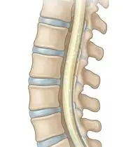
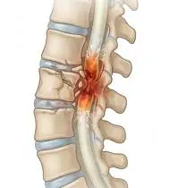
Spinal Cord Injury (SCI) — Overview
Spinal Cord Injury (SCI) is a traumatic or non-traumatic lesion of the neural elements within the spinal canal resulting in partial (Incomplete: ASIA B, C, D) or total (Complete: ASIA A) loss of motor, sensory, and autonomic function below the Neurological Level of Injury (NLI — the most caudal segment with antigravity ≥3/5 motor strength [provided the rostral segment is 5/5] and intact 2/2 pinprick & light touch sensation bilaterally). Clinically, acute SCI begins with Spinal Shock (complete flaccid paralysis, areflexia, and loss of bulbocavernosus reflex for days to weeks), followed by emergence of spasticity, hyperreflexia, neurogenic bladder/bowel, and in lesions at or above T6, life-threatening Autonomic Dysreflexia!
A Spinal Cord Injury (SCI) is damage to the main nerve highway inside the spine that carries movement and sensation signals between the brain and body. Whether the injury is incomplete (some signals still pass through and can be strengthened through neuroplasticity) or complete (focusing on upper-body power, transfers, and independent mobility), proactive physiotherapy protects your lungs, skin, joints, and functional freedom!
Primary Causes & Biomechanics
Key Points You Should Know
- •Traumatic vertebral fracture-dislocation (motor vehicle crashes, falls, diving into shallow water) or non-traumatic ischemia/myelitis/tumor
- •ISNCSCI (ASIA Impairment Scale): ASIA A (Complete: no sacral S4–S5 motor/sensory preservation) vs. ASIA B–D (Incomplete: Sacral Sparing present!—voluntary anal contraction or S4–S5 perianal sensation indicates intact long tracts and vastly superior recovery prognosis!)
- •Systemic SCI Secondary Complications: Autonomic Dysreflexia (≥T6), Orthostatic Hypotension, Pressure Ulcers (ischial/sacral), DVT, Heterotopic Ossification (hips/knees), and Respiratory Atelectasis
- •The 20-Minute Pressure-Relief Rule (Skin Protection): When sensation is reduced below the injury, sitting in one spot without shifting weight cuts off blood flow to the skin over your sit-bones! Doing a 15–30 second wheelchair push-up (or leaning side-to-side/forward) every 20–30 minutes prevents dangerous pressure sores.
- •Recognizing Autonomic Dysreflexia (For Injuries at T6 and Above — Emergency!): A sudden pounding headache, sweating/flushing above the injury, and goosebumps mean your blood pressure is spiking—usually caused by a full bladder (kinked catheter) or tight clothing. Sit upright immediately (drop your legs down) and check your catheter!
- •Gentle Daily Stretching Prevents Hip & Ankle Stiffness: Moving the legs smoothly every day keeps joints flexible for standing frames, braces, and dressing.
Assessment Methods
1. Goniometric ROM & Postural / Kinematic Analysis
ISNCSCI (ASIA) Examination: 10 Key Myotomes (C5 Elbow Flexors, C6 Wrist Extensors, C7 Elbow Extensors, C8 Finger Flexors, T1 Small Finger Abductors; L2 Hip Flexors, L3 Knee Extensors, L4 Ankle Dorsiflexors, L5 Long Toe Extensors, S1 Ankle Plantarflexors) and 28 Dermatomes (C2–S4/5).
2. Special Clinical Provocation & Neurological/Functional Tests
Modified Ashworth Scale (spasticity), SCIM-III (Spinal Cord Independence Measure), WISCI-II (Walking Index for SCI), Forced Vital Capacity (FVC), and daily skin pressure inspection.
3. Standardized Clinical Outcome Scales & Imaging Criteria
ASIA Impairment Scale (A–E), SCIM-III, WISCI-II, and spine MRI/CT.
1. Range of Motion & Posture Check
Your physiotherapist gently evaluates how comfortably and smoothly your spinal cord (ascending sensory tracts, descending corticospinal/autonomic tracts, and anterior horn motor neurons) and vertebral column moves and checks your overall posture.
2. Muscle Strength & Functional Movement Check
Your muscle balance, walking or reaching comfort, and specific gentle movement tests are evaluated without triggering sharp pain.
3. Typical Symptoms & Daily Impact Review
Loss or reduction of voluntary muscle strength and skin sensation (touch, pain, temperature) below the spinal segment of injury, accompanied by neurogenic bladder and bowel dysfunction.
Treatment Options & Phase-Based Protocol
✨ Enriched GIF Exercise & Precaution GuidePhase 1: Acute Symptom Modulation, Tissue Protection & Early Activation
- •Phase 1 Clinical Focus & Tissue Protection: Phase 1 — Acute / Spinal Shock & Prevention Phase: 2-hourly turning & pressure-relief positioning, daily passive/active-assisted ROM across all joints (ankle pumps, towel gastro-soleus stretches, supine heel slides, wand shoulder elevation) to prevent contractures and Heterotopic Ossification, and diaphragmatic/thoracic expansion breathing + assisted cough.
- •Pain, Edema & Neuromuscular Modulation: Targeted physical modalities, relative load management, and early protected mobility of the spinal cord (ascending sensory tracts, descending corticospinal/autonomic tracts, and anterior horn motor neurons) and vertebral column without provoking mechanical or inflammatory irritation.
- •Proximal & Distal Kinetic Chain Maintenance: Maintain active mobility and circulation in adjacent joints while protecting the primary healing structures in Spinal Cord Injury.
Phase 2: Progressive Mobility, Motor Control & Targeted Strengthening
- •Phase 2 Progressive Mobilization & Motor Control: Phase 2 — Tilt-Table / Seated Balance, Upper Limb & Transfer Phase: Gradual upright tilt-table/reclining wheelchair progression (with abdominal binder & compression stockings for orthostatic hypotension!), seated wheelchair push-ups (30s pressure relief every 20 minutes!), and scapular/triceps/latissimus strengthening.
- •Biomechanical Alignment & Co-Contraction: Restore balanced agonist-antagonist force couples, joint centration, and multi-planar neuromuscular control across the spinal cord (ascending sensory tracts, descending corticospinal/autonomic tracts, and anterior horn motor neurons) and vertebral column.
- •Myofascial Extensibility & Endurance: Address secondary compensatory tightness and build local muscular endurance within safe, symptom-free biomechanical parameters.
Phase 3: Functional Integration, Proprioception & Long-Term Prevention
- •Phase 3 Functional Integration & Long-Term Prevention: Phase 3 — Incomplete Neuroplastic Gait or Wheelchair Independence Phase: In Incomplete (ASIA C/D) injuries, perform pelvic bridging, sit-to-stand, side-lying hip abduction, and Body-Weight-Supported Treadmill / KAFO-AFO gait training; in Complete lesions, master independent bed-to-wheelchair transfers and community mobility.
- •Proprioceptive Balance & Dynamic Loading: Progress to task-specific closed-chain, balance, and functional movement patterns tailored to daily life, occupational, or athletic demands.
- •Self-Management & Relapse Prophylaxis: Establish lifelong ergonomic habits, home exercise maintenance, and early warning-sign awareness to prevent recurrence.
Step 1: Calming Pain, Protecting the Area & Gentle Starter Motion
- •The 20-Minute Pressure-Relief Rule (Skin Protection): When sensation is reduced below the injury, sitting in one spot without shifting weight cuts off blood flow to the skin over your sit-bones! Doing a 15–30 second wheelchair push-up (or leaning side-to-side/forward) every 20–30 minutes prevents dangerous pressure sores.
- •Calm Protection & Gentle Start: During the initial stage of Spinal Cord Injury / Paralysis, avoid painful overloading and perform the gentle, pain-free starter movements shown below to support circulation and comfort.
- •Respecting Your Body's Signals: Every movement should stay within a comfortable, non-painful zone—never force through sharp or radiating pain.
Step 2: Restoring Flexibility & Building Supporting Muscle Strength
- •Recognizing Autonomic Dysreflexia (For Injuries at T6 and Above — Emergency!): A sudden pounding headache, sweating/flushing above the injury, and goosebumps mean your blood pressure is spiking—usually caused by a full bladder (kinked catheter) or tight clothing. Sit upright immediately (drop your legs down) and check your catheter!
- •Step-by-Step Flexibility & Support: As acute sensitivity settles, gradually build mobility and strengthen the supporting muscles around the joint at a steady, controlled pace.
- •Posture & Breathing Coordination: Keep your posture upright and breathe smoothly (never hold your breath!) during every repetition.
Step 3: Safe Daily Habits, Stamina & Preventing Recurrence
- •Gentle Daily Stretching Prevents Hip & Ankle Stiffness: Moving the legs smoothly every day keeps joints flexible for standing frames, braces, and dressing.
- •Confident Return to Daily Life: Integrate your stronger muscles and improved balance into walking, stairs, work, and recreational activities.
- •Preventing Future Flare-Ups: Continue your core protective habits and key maintenance exercises even after symptoms resolve.
Animated (GIF) Therapeutic Exercise Guide
Animated clinical exercise protocol with biomechanical angles, target muscle groups, and dosage parameters (Click image to enlarge)
Step-by-step animated exercise demonstrations you can safely perform at home (Click image to enlarge)
 🔍 Enlarge
1. Supine Active-Assisted Heel Slide & Synovial Diffusion Mobilization
1. Supine Heel Slide (Gentle Knee & Hip Bending)
Controlled flexion and extension of the lower extremity by sliding the heel along the support surface without axial weight-bearing. Stimulates synovial fluid diffusion across avascular cartilage and prevents capsular adhesions. Specifically indicated in Spinal Cord Injury rehabilitation to optimize spinal cord (ascending sensory tracts, descending corticospinal/autonomic tracts, and anterior horn motor neurons) and vertebral column biomechanics and neuromuscular control.
While lying on your back, slowly slide your heel along the bed sheet toward your hip within a comfortable, pain-free angle, hold for 3 seconds, and gently slide it back out straight. Perform smoothly within a comfortable, pain-free range to support your Spinal Cord Injury / Paralysis recovery.
 🔍 Enlarge
2. Rhythmic Active Ankle Dorsiflexion & Plantarflexion Pumping (Venous/Edema Pump)
2. Ankle Toe-Pulls and Pointing (Circulation & Swelling Pump)
Rhythmic alternating contraction of the tibialis anterior and gastrocnemius-soleus complex. Activates the deep venous calf pump to prevent deep vein thrombosis (DVT), clears distal edema, and maintains talocrural mobility. Specifically indicated in Spinal Cord Injury rehabilitation to optimize spinal cord (ascending sensory tracts, descending corticospinal/autonomic tracts, and anterior horn motor neurons) and vertebral column biomechanics and neuromuscular control.
While lying down or sitting with your legs supported, pull your toes and feet firmly upward toward your shins, hold for 3 seconds, then point them downward like pressing a gas pedal. Perform smoothly within a comfortable, pain-free range to support your Spinal Cord Injury / Paralysis recovery.
 🔍 Enlarge
3. Seated Depression Transfer Lift ('Push-Up' — Triceps, Latissimus & Pressure Relief)
3. Seated Arm-Press Trunk Lift (Transfer & Pressure Relief)
Pressing down on armrests or push-blocks with elbows extending and scapulae depressing to lift the buttocks clear of the seat. Essential for independent bed-to-wheelchair transfers, crutch ambulation, and relieving ischial tuberosity pressure every 20–30 minutes. Specifically indicated in Spinal Cord Injury rehabilitation to optimize spinal cord (ascending sensory tracts, descending corticospinal/autonomic tracts, and anterior horn motor neurons) and vertebral column biomechanics and neuromuscular control.
While sitting securely in your chair or wheelchair with brakes locked, place your hands on the armrests and push down to lift your hips slightly off the seat for 5–10 seconds, then lower gently. Perform smoothly within a comfortable, pain-free range to support your Spinal Cord Injury / Paralysis recovery.
 🔍 Enlarge
4. Scapulothoracic Retraction & Depression (Middle/Lower Trapezius & Rhomboids)
4. Shoulder Blade Squeeze (Postural Alignment)
Adduction and depression of the scapulae via the middle/lower trapezius and rhomboids while inhibiting upper trapezius overactivity. Corrects anterior scapular tilt, widens the subacromial outlet, and unloads the anterior thoracic column. Specifically indicated in Spinal Cord Injury rehabilitation to optimize spinal cord (ascending sensory tracts, descending corticospinal/autonomic tracts, and anterior horn motor neurons) and vertebral column biomechanics and neuromuscular control.
Sit or stand tall. Keep your shoulders relaxed away from your ears, gently draw your shoulder blades together and slightly downward toward your back pockets, hold for 5 seconds, and release. Perform smoothly within a comfortable, pain-free range to support your Spinal Cord Injury / Paralysis recovery.
 🔍 Enlarge
5. Thoracic Expansion Exercise (TEE) with Bilateral Arm Elevation & 3s Inspiratory Hold
5. Chest-Opening Deep Breath with Arm Raise
Synchronizing deep nasal inspiration with bilateral shoulder flexion/abduction and a 3-second end-inspiratory hold before passive/pursed-lip expiration. Opens collateral ventilation channels to prevent basal atelectasis and maintains costovertebral compliance. Specifically indicated in Spinal Cord Injury rehabilitation to optimize spinal cord (ascending sensory tracts, descending corticospinal/autonomic tracts, and anterior horn motor neurons) and vertebral column biomechanics and neuromuscular control.
While sitting upright, breathe in deeply through your nose while opening your arms upward and outward to expand your ribcage. Hold the breath inside for 3 seconds, then exhale slowly while lowering your arms. Perform smoothly within a comfortable, pain-free range to support your Spinal Cord Injury / Paralysis recovery.
 🔍 Enlarge
6. Prone Positioning & Controlled Thoracolumbar Extension (McKenzie / Back Extensor)
6. Prone Lying & Gentle Back Extension Lift
Prone positioning and graduated thoracic/lumbar extension (with a pillow under the abdomen when indicated for spinal unloading). Counters flexed postural kyphosis, stretches tight hip flexors, and strengthens paraspinal extensors. Specifically indicated in Spinal Cord Injury rehabilitation to optimize spinal cord (ascending sensory tracts, descending corticospinal/autonomic tracts, and anterior horn motor neurons) and vertebral column biomechanics and neuromuscular control.
Lie on your stomach (place a small pillow under your hips/tummy if needed for comfort). Gently lift your head and chest slightly off the mat by squeezing your back muscles without straining your neck, hold for 3–5 seconds, and lower. Perform smoothly within a comfortable, pain-free range to support your Spinal Cord Injury / Paralysis recovery.
Movements to Avoid & Strict Precautions
1. Sitting in a Wheelchair for >30 Minutes Without Performing a Pressure-Relief Lift/Lean
1. Sitting for more than 20–30 minutes without lifting or leaning to relieve pressure on your sit-bones
2. Lying Flat When Experiencing a Pounding Headache in a T6+ Injury (Worsens Brain Blood Pressure in Autonomic Dysreflexia!)
2. Lying flat during an Autonomic Dysreflexia headache (ALWAYS sit upright immediately to lower blood pressure!)
3. Forceful Passive Stretching of Insensate Hips or Knees (Causes Heterotopic Bone Growth or Fractures!)
3. Forcefully wrenching stiff hips or knees that lack pain sensation
4. Placing Hot Water Bottles or Heating Pads on Areas With Reduced Sensation (Causes Severe Burns!)
4. Putting hot packs or hot water bottles on skin that cannot feel temperature
Spinal Cord Injury (SCI) — Video Guide
Click directly on any video to open and watch in a window, or review details via 📋 Summary & Options.
Condition Overview Video (1 Video)
Summary of pathophysiology, biomechanics, and clinical presentation
What the condition is, why it develops, and its basic mechanism

What Is a Spinal Cord Injury (SCI)? Neurological Levels and Clinical Presentation
Spinal Cord Injury (SCI) is a traumatic or non-traumatic lesion of the neural elements within the spinal canal resulting in partial (Incomplete: ASIA B, C, D) or total (Complete: ASIA A) loss of motor, sensory, and autonomic function below the Neurological Level of Injury (NLI — the most caudal segment with antigravity ≥3/5 motor strength [provided the rostral segment is 5/5] and intact 2/2 pinprick & light touch sensation bilaterally). Clinically, acute SCI begins with Spinal Shock (complete flaccid paralysis, areflexia, and loss of bulbocavernosus reflex for days to weeks), followed by emergence of spasticity, hyperreflexia, neurogenic bladder/bowel, and in lesions at or above T6, life-threatening Autonomic Dysreflexia!
A Spinal Cord Injury (SCI) is damage to the main nerve highway inside the spine that carries movement and sensation signals between the brain and body. Whether the injury is incomplete (some signals still pass through and can be strengthened through neuroplasticity) or complete (focusing on upper-body power, transfers, and independent mobility), proactive physiotherapy protects your lungs, skin, joints, and functional freedom!
Treatment and Exercise Videos
Phase-based physiotherapy protocols, mobilization, and therapeutic exercises
Step-by-step recovery and safe home exercise applications

Home Exercises and Transfer Training for Spinal Cord Injury (Paraplegia / Tetraplegia)
Phase 1: Acute Symptom Modulation, Tissue Protection & Early Activation: Phase 1 Clinical Focus & Tissue Protection: Phase 1 — Acute / Spinal Shock & Prevention Phase: 2-hourly turning & pressure-relief positioning, daily passive/active-assisted ROM across all joints (ankle pumps, towel gastro-soleus stretches, supine heel slides, wand shoulder elevation) to prevent contractures and Heterotopic Ossification, and diaphragmatic/thoracic expansion breathing + assisted cough.
Step 1: Calming Pain, Protecting the Area & Gentle Starter Motion: The 20-Minute Pressure-Relief Rule (Skin Protection): When sensation is reduced below the injury, sitting in one spot without shifting weight cuts off blood flow to the skin over your sit-bones! Doing a 15–30 second wheelchair push-up (or leaning side-to-side/forward) every 20–30 minutes prevents dangerous pressure sores.

Seated Balance and Trunk Strengthening Exercises After Spinal Cord Injury
Phase 2: Progressive Mobility, Motor Control & Targeted Strengthening: Phase 2 Progressive Mobilization & Motor Control: Phase 2 — Tilt-Table / Seated Balance, Upper Limb & Transfer Phase: Gradual upright tilt-table/reclining wheelchair progression (with abdominal binder & compression stockings for orthostatic hypotension!), seated wheelchair push-ups (30s pressure relief every 20 minutes!), and scapular/triceps/latissimus strengthening.
Step 2: Restoring Flexibility & Building Supporting Muscle Strength: Recognizing Autonomic Dysreflexia (For Injuries at T6 and Above — Emergency!): A sudden pounding headache, sweating/flushing above the injury, and goosebumps mean your blood pressure is spiking—usually caused by a full bladder (kinked catheter) or tight clothing. Sit upright immediately (drop your legs down) and check your catheter!
Clinical Assessment, Goniometry & Special Test Videos
Displayed only in Healthcare Professional mode (hidden in Patient mode)

Systematic Neurological Examination: Reflex, Sensory, Tone, and Motor Assessment
Goniometric ROM & Postural / Kinematic Analysis: ISNCSCI (ASIA) Examination: 10 Key Myotomes (C5 Elbow Flexors, C6 Wrist Extensors, C7 Elbow Extensors, C8 Finger Flexors, T1 Small Finger Abductors; L2 Hip Flexors, L3 Knee Extensors, L4 Ankle Dorsiflexors, L5 Long Toe Extensors, S1 Ankle Plantarflexors) and 28 Dermatomes (C2–S4/5).

Manual Muscle Testing (MRC 0–5 Grading) — Clinical Motor Strength Examination
Special Clinical Provocation & Neurological/Functional Tests: Modified Ashworth Scale (spasticity), SCIM-III (Spinal Cord Independence Measure), WISCI-II (Walking Index for SCI), Forced Vital Capacity (FVC), and daily skin pressure inspection.

Modified Ashworth Scale (MAS) — Clinical Spasticity Assessment
Standardized Clinical Outcome Scales & Imaging Criteria: ASIA Impairment Scale (A–E), SCIM-III, WISCI-II, and spine MRI/CT.