Hemiplegia (Post-Stroke Rehabilitation)Stroke / Partial Paralysis (Hemiplegia)
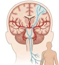
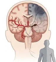
Hemiplegia (Post-Stroke Rehabilitation) — Overview
Hemiplegia (post-stroke / cerebrovascular accident [CVA] hemiparesis) is an upper motor neuron (UMN) syndrome affecting the contralateral half of the body following ischemic (~80–85%, e.g., Middle Cerebral Artery infarction affecting upper limb/face > lower limb, or Anterior Cerebral Artery affecting lower limb > upper limb) or hemorrhagic (~15–20%) brain injury. Motor recovery classically progresses through Brunnstrom's 6 Stages: Stage 1 Flaccidity -> Stage 2 Spasticity & Associated Reactions appear -> Stage 3 Peak Spasticity & Stereotypic Flexor/Extensor Synergies -> Stage 4 Movement Deviating from Synergy -> Stage 5 Selective Independent Movement -> Stage 6 Near-Normal Coordination. Rehabilitation leverages task-specific neuroplasticity, Bobath/NDT postural control, and strict hemiplegic shoulder protection.
Hemiplegia (post-stroke partial paralysis) occurs when a blocked or bleeding blood vessel in one side of the brain affects the movement and balance centers for the opposite side of the body (a right-brain stroke affects the left arm and leg; a left-brain stroke affects the right arm and leg). Because the brain has an amazing ability to rewire itself (neuroplasticity), consistent, purposeful movement re-teaches healthy brain pathways to take over!
Primary Causes & Biomechanics
Key Points You Should Know
- •Ischemic thromboembolism or intracerebral hemorrhage disrupting corticospinal and reticulospinal descending pathways
- •Loss of cortical inhibition releasing velocity-dependent tonic stretch reflexes (spasticity) and stereotypic Upper Limb Flexor Synergy (scapular retraction/depression, shoulder adduction/IR, elbow flexion, forearm pronation, wrist/finger flexion) & Lower Limb Extensor Synergy (hip extension/adduction/IR, knee extension, ankle plantarflexion/inversion — causing circumduction gait)
- •Hemiplegic Glenohumeral Subluxation & Shoulder-Hand Syndrome (CRPS Type I) during the early flaccid stage if the paralyzed arm is pulled or left unsupported!
- •Golden Rule #1 — Protect the Paralyzed Shoulder: In the early weeks when the shoulder muscles are loose, NEVER pull on the weak arm to help the patient sit up or roll over, and always keep a pillow supporting the elbow when sitting! Pulling on a weak arm causes painful shoulder subluxation.
- •Golden Rule #2 — NEVER Squeeze a Hard Rubber Ball! In stroke recovery, the hand naturally wants to curl into a tight fist (spasticity). Squeezing a stress ball makes the hand curl tighter! Instead, practice opening the fingers flat on a table and lifting the wrist upward.
- •Loading the Weak Leg When Standing Up: When rising from a chair, keep the weak foot slightly back and press evenly through *both* feet so the brain doesn't 'forget' the affected leg.
Assessment Methods
1. Goniometric ROM & Postural / Kinematic Analysis
Brunnstrom Motor Recovery Stages (1–6) for hand, upper extremity, and lower extremity; Modified Ashworth Scale (MAS 0–4) for spasticity; and Fugl-Meyer Assessment (FMA).
2. Special Clinical Provocation & Neurological/Functional Tests
Berg Balance Scale (BBS), Trunk Impairment Scale (TIS), Functional Ambulation Category (FAC 0–5), glenohumeral subluxation sulcus measurement (in fingerbreadths), and screening for left spatial neglect (in right parietal stroke) and aphasia/dysphagia.
3. Standardized Clinical Outcome Scales & Imaging Criteria
Fugl-Meyer Assessment, Barthel Index / FIM, 10-Meter Walk Test, and Timed Up and Go (TUG).
1. Range of Motion & Posture Check
Your physiotherapist gently evaluates how comfortably and smoothly your cerebral motor cortex, internal capsule, corticospinal (pyramidal) tract, and contralateral hemibody moves and checks your overall posture.
2. Muscle Strength & Functional Movement Check
Your muscle balance, walking or reaching comfort, and specific gentle movement tests are evaluated without triggering sharp pain.
3. Typical Symptoms & Daily Impact Review
Unilateral weakness or paralysis (hemiparesis/hemiplegia) of the arm, leg, trunk, and lower face on the side opposite the brain lesion.
Treatment Options & Phase-Based Protocol
✨ Enriched GIF Exercise & Precaution GuidePhase 1: Acute Symptom Modulation, Tissue Protection & Early Activation
- •Phase 1 Clinical Focus & Tissue Protection: Phase 1 — Acute / Flaccid-to-Early Synergy Phase (Brunnstrom 1–2): Anti-spastic bed positioning (scapula protracted on a pillow, shoulder abducted/externally rotated, elbow/wrist/fingers extended), NEVER pull on the hemiplegic arm during transfers!, Bobath bilateral clasped-hand elevation (affected thumb on top, elbows straight), and supine heel slides & pelvic bridging (breaking lower limb extensor synergy!).
- •Pain, Edema & Neuromuscular Modulation: Targeted physical modalities, relative load management, and early protected mobility of the cerebral motor cortex, internal capsule, corticospinal (pyramidal) tract, and contralateral hemibody without provoking mechanical or inflammatory irritation.
- •Proximal & Distal Kinetic Chain Maintenance: Maintain active mobility and circulation in adjacent joints while protecting the primary healing structures in Hemiplegia.
Phase 2: Progressive Mobility, Motor Control & Targeted Strengthening
- •Phase 2 Progressive Mobilization & Motor Control: Phase 2 — Spasticity Modulation, Trunk Control & Weight-Bearing Phase (Brunnstrom 3–4): Slow sustained towel gastro-soleus & wrist/finger flexor stretching (inhibiting spasticity without squeezing rubber balls!), active finger spreading & wrist extension, side-lying hip abduction, and symmetrical chair sit-to-stand transfers (loading the affected foot!).
- •Biomechanical Alignment & Co-Contraction: Restore balanced agonist-antagonist force couples, joint centration, and multi-planar neuromuscular control across the cerebral motor cortex, internal capsule, corticospinal (pyramidal) tract, and contralateral hemibody.
- •Myofascial Extensibility & Endurance: Address secondary compensatory tightness and build local muscular endurance within safe, symptom-free biomechanical parameters.
Phase 3: Functional Integration, Proprioception & Long-Term Prevention
- •Phase 3 Functional Integration & Long-Term Prevention: Phase 3 — Selective Motor Control, Balance & Gait Re-Education Phase (Brunnstrom 5–6): Supported weight-shifting & tandem balance, overcoming knee hyperextension (genu recurvatum) and foot drop (with AFO or FES if needed), Constraint-Induced Movement Therapy (CIMT) or bimanual task training.
- •Proprioceptive Balance & Dynamic Loading: Progress to task-specific closed-chain, balance, and functional movement patterns tailored to daily life, occupational, or athletic demands.
- •Self-Management & Relapse Prophylaxis: Establish lifelong ergonomic habits, home exercise maintenance, and early warning-sign awareness to prevent recurrence.
Step 1: Calming Pain, Protecting the Area & Gentle Starter Motion
- •Golden Rule #1 — Protect the Paralyzed Shoulder: In the early weeks when the shoulder muscles are loose, NEVER pull on the weak arm to help the patient sit up or roll over, and always keep a pillow supporting the elbow when sitting! Pulling on a weak arm causes painful shoulder subluxation.
- •Calm Protection & Gentle Start: During the initial stage of Stroke / Partial Paralysis, avoid painful overloading and perform the gentle, pain-free starter movements shown below to support circulation and comfort.
- •Respecting Your Body's Signals: Every movement should stay within a comfortable, non-painful zone—never force through sharp or radiating pain.
Step 2: Restoring Flexibility & Building Supporting Muscle Strength
- •Golden Rule #2 — NEVER Squeeze a Hard Rubber Ball! In stroke recovery, the hand naturally wants to curl into a tight fist (spasticity). Squeezing a stress ball makes the hand curl tighter! Instead, practice opening the fingers flat on a table and lifting the wrist upward.
- •Step-by-Step Flexibility & Support: As acute sensitivity settles, gradually build mobility and strengthen the supporting muscles around the joint at a steady, controlled pace.
- •Posture & Breathing Coordination: Keep your posture upright and breathe smoothly (never hold your breath!) during every repetition.
Step 3: Safe Daily Habits, Stamina & Preventing Recurrence
- •Loading the Weak Leg When Standing Up: When rising from a chair, keep the weak foot slightly back and press evenly through *both* feet so the brain doesn't 'forget' the affected leg.
- •Confident Return to Daily Life: Integrate your stronger muscles and improved balance into walking, stairs, work, and recreational activities.
- •Preventing Future Flare-Ups: Continue your core protective habits and key maintenance exercises even after symptoms resolve.
Animated (GIF) Therapeutic Exercise Guide
Animated clinical exercise protocol with biomechanical angles, target muscle groups, and dosage parameters (Click image to enlarge)
Step-by-step animated exercise demonstrations you can safely perform at home (Click image to enlarge)
 🔍 Enlarge
1. Bobath Bilateral Clasp-Hand Elevation & Midline Trunk Organization
1. Clasped-Hands Two-Arm Overhead Reach
Interlocking the fingers with the affected thumb positioned on top, maintaining elbow extension and scapular protraction while elevating both arms symmetrically in the midline. Inhibits upper limb flexor synergy and prevents hemiplegic shoulder impingement. Specifically indicated in Hemiplegia rehabilitation to optimize cerebral motor cortex, internal capsule, corticospinal (pyramidal) tract, and contralateral hemibody biomechanics and neuromuscular control.
Clasp your hands together with your affected thumb on top. Keeping both elbows straight, use your stronger arm to smoothly guide both hands upward toward the ceiling and slowly back down. Perform smoothly within a comfortable, pain-free range to support your Stroke / Partial Paralysis recovery.
 🔍 Enlarge
2. Dorsal/Palmar Interossei & Lumbrical Activation (Active Finger Abduction/Extension)
2. Tabletop Finger Spreading (Fan Out) & Opening
With the palm resting flat on a surface, actively spreading all digits apart (abduction) and bringing them back together, combined with controlled MCP/IP extension. Strengthens ulnar-innervated interossei and balances extrinsic flexors. Specifically indicated in Hemiplegia rehabilitation to optimize cerebral motor cortex, internal capsule, corticospinal (pyramidal) tract, and contralateral hemibody biomechanics and neuromuscular control.
Rest your hand flat on a table. Spread your fingers wide apart like a fan, hold for 3 seconds, and slowly bring them back together without lifting your palm. Perform smoothly within a comfortable, pain-free range to support your Stroke / Partial Paralysis recovery.
 🔍 Enlarge
3. Supine Active-Assisted Heel Slide & Synovial Diffusion Mobilization
3. Supine Heel Slide (Gentle Knee & Hip Bending)
Controlled flexion and extension of the lower extremity by sliding the heel along the support surface without axial weight-bearing. Stimulates synovial fluid diffusion across avascular cartilage and prevents capsular adhesions. Specifically indicated in Hemiplegia rehabilitation to optimize cerebral motor cortex, internal capsule, corticospinal (pyramidal) tract, and contralateral hemibody biomechanics and neuromuscular control.
While lying on your back, slowly slide your heel along the bed sheet toward your hip within a comfortable, pain-free angle, hold for 3 seconds, and gently slide it back out straight. Perform smoothly within a comfortable, pain-free range to support your Stroke / Partial Paralysis recovery.
 🔍 Enlarge
4. Supine Lumbopelvic Bridging (Gluteus Maximus & Posterior Chain Co-Contraction)
4. Supine Hip Bridge (Controlled Pelvic Lift)
From hook-lying with transversus abdominis pre-activation, elevating the pelvis until hips, pelvis, and shoulders form a neutral diagonal line. Strengthens the gluteus maximus, hamstrings, and erector spinae synergy without lumbar hyperlordosis. Specifically indicated in Hemiplegia rehabilitation to optimize cerebral motor cortex, internal capsule, corticospinal (pyramidal) tract, and contralateral hemibody biomechanics and neuromuscular control.
Lie on your back with your knees bent and feet flat hip-width apart. Gently tighten your tummy and glutes, lift your hips until your body forms a straight line from shoulders to knees, hold for 5 seconds, and lower slowly. Perform smoothly within a comfortable, pain-free range to support your Stroke / Partial Paralysis recovery.
 🔍 Enlarge
5. Functional Closed-Kinetic-Chain Chair Transfer (Sit-to-Stand Biomechanics)
5. Controlled Chair Sit-to-Stand Transfer
Controlled transition from sitting to standing and eccentric lowering back to the chair with symmetrical weight distribution, anterior trunk inclination ('nose over toes'), and neutral frontal knee alignment. Specifically indicated in Hemiplegia rehabilitation to optimize cerebral motor cortex, internal capsule, corticospinal (pyramidal) tract, and contralateral hemibody biomechanics and neuromuscular control.
Sit near the front of a sturdy chair with your feet hip-width apart. Lean slightly forward and push evenly through both heels to stand up tall, then slowly lower yourself back down over 3 seconds. Perform smoothly within a comfortable, pain-free range to support your Stroke / Partial Paralysis recovery.
 🔍 Enlarge
6. Single-Leg / Semi-Tandem Proprioceptive & Weight-Shift Balance Training
6. Supported Weight-Shifting & Tandem Balance Practice
Controlled mediolateral/anteroposterior weight shifting and tandem or single-leg stance near a stable support. Enhances mechanoreceptor feedback, ankle/hip postural reflexes, and dynamic gait stability. Specifically indicated in Hemiplegia rehabilitation to optimize cerebral motor cortex, internal capsule, corticospinal (pyramidal) tract, and contralateral hemibody biomechanics and neuromuscular control.
Stand near a counter or sturdy chair for safety. Place one foot slightly in front of the other (or shift your weight smoothly from side to side) and hold your balance upright for 20–30 seconds. Perform smoothly within a comfortable, pain-free range to support your Stroke / Partial Paralysis recovery.
Movements to Avoid & Strict Precautions
1. Pulling on the Weak/Paralyzed Arm During Bed Rolling or Chair Transfers (Dislocates the Shoulder!)
1. NEVER pull on the patient's weak arm to help them sit up, turn in bed, or walk (always support under the shoulder blade and trunk!)
2. Squeezing a Stress Ball or Hand-Gripper (Reinforces Flexor Spasticity and Closes the Hand Tighter!)
2. Squeezing a rubber ball in the weak hand (makes finger curling spasticity worse—always practice opening the hand instead!)
3. Letting the Paralyzed Arm Hang Limply Down Beside the Wheelchair Without Pillow/Lap-Tray Support
3. Letting the weak arm dangle unsupported beside a chair or bed
4. Pushing Up Only With the Strong Leg While Ignoring the Affected Side ('Learned Non-Use')
4. Doing everything only with the strong side and ignoring the weak arm and leg
Hemiplegia (Post-Stroke Rehabilitation) — Video Guide
Click directly on any video to open and watch in a window, or review details via 📋 Summary & Options.
Condition Overview Video (1 Video)
Summary of pathophysiology, biomechanics, and clinical presentation
What the condition is, why it develops, and its basic mechanism

What Is Hemiplegia (Stroke / Paralysis)? Causes and Clinical Presentation
Hemiplegia (post-stroke / cerebrovascular accident [CVA] hemiparesis) is an upper motor neuron (UMN) syndrome affecting the contralateral half of the body following ischemic (~80–85%, e.g., Middle Cerebral Artery infarction affecting upper limb/face > lower limb, or Anterior Cerebral Artery affecting lower limb > upper limb) or hemorrhagic (~15–20%) brain injury. Motor recovery classically progresses through Brunnstrom's 6 Stages: Stage 1 Flaccidity -> Stage 2 Spasticity & Associated Reactions appear -> Stage 3 Peak Spasticity & Stereotypic Flexor/Extensor Synergies -> Stage 4 Movement Deviating from Synergy -> Stage 5 Selective Independent Movement -> Stage 6 Near-Normal Coordination. Rehabilitation leverages task-specific neuroplasticity, Bobath/NDT postural control, and strict hemiplegic shoulder protection.
Hemiplegia (post-stroke partial paralysis) occurs when a blocked or bleeding blood vessel in one side of the brain affects the movement and balance centers for the opposite side of the body (a right-brain stroke affects the left arm and leg; a left-brain stroke affects the right arm and leg). Because the brain has an amazing ability to rewire itself (neuroplasticity), consistent, purposeful movement re-teaches healthy brain pathways to take over!
Treatment and Exercise Videos
Phase-based physiotherapy protocols, mobilization, and therapeutic exercises
Step-by-step recovery and safe home exercise applications
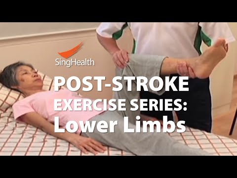
Hemiplegia (Post-Stroke) Physical Therapy and Functional Exercises
Phase 1: Acute Symptom Modulation, Tissue Protection & Early Activation: Phase 1 Clinical Focus & Tissue Protection: Phase 1 — Acute / Flaccid-to-Early Synergy Phase (Brunnstrom 1–2): Anti-spastic bed positioning (scapula protracted on a pillow, shoulder abducted/externally rotated, elbow/wrist/fingers extended), NEVER pull on the hemiplegic arm during transfers!, Bobath bilateral clasped-hand elevation (affected thumb on top, elbows straight), and supine heel slides & pelvic bridging (breaking lower limb extensor synergy!).
Step 1: Calming Pain, Protecting the Area & Gentle Starter Motion: Golden Rule #1 — Protect the Paralyzed Shoulder: In the early weeks when the shoulder muscles are loose, NEVER pull on the weak arm to help the patient sit up or roll over, and always keep a pillow supporting the elbow when sitting! Pulling on a weak arm causes painful shoulder subluxation.

Bobath Auto-Inhibition and Spasticity Control in Stroke (Hemiplegia) Rehabilitation
Phase 2: Progressive Mobility, Motor Control & Targeted Strengthening: Phase 2 Progressive Mobilization & Motor Control: Phase 2 — Spasticity Modulation, Trunk Control & Weight-Bearing Phase (Brunnstrom 3–4): Slow sustained towel gastro-soleus & wrist/finger flexor stretching (inhibiting spasticity without squeezing rubber balls!), active finger spreading & wrist extension, side-lying hip abduction, and symmetrical chair sit-to-stand transfers (loading the affected foot!).
Step 2: Restoring Flexibility & Building Supporting Muscle Strength: Golden Rule #2 — NEVER Squeeze a Hard Rubber Ball! In stroke recovery, the hand naturally wants to curl into a tight fist (spasticity). Squeezing a stress ball makes the hand curl tighter! Instead, practice opening the fingers flat on a table and lifting the wrist upward.
Clinical Assessment, Goniometry & Special Test Videos
Displayed only in Healthcare Professional mode (hidden in Patient mode)

Systematic Neurological Examination: Reflex, Sensory, Tone, and Motor Assessment
Goniometric ROM & Postural / Kinematic Analysis: Brunnstrom Motor Recovery Stages (1–6) for hand, upper extremity, and lower extremity; Modified Ashworth Scale (MAS 0–4) for spasticity; and Fugl-Meyer Assessment (FMA).

Modified Ashworth Scale (MAS) — Clinical Spasticity Assessment
Special Clinical Provocation & Neurological/Functional Tests: Berg Balance Scale (BBS), Trunk Impairment Scale (TIS), Functional Ambulation Category (FAC 0–5), glenohumeral subluxation sulcus measurement (in fingerbreadths), and screening for left spatial neglect (in right parietal stroke) and aphasia/dysphagia.

Berg Balance Scale (BBS) — Clinical Postural Control and Balance Examination
Standardized Clinical Outcome Scales & Imaging Criteria: Fugl-Meyer Assessment, Barthel Index / FIM, 10-Meter Walk Test, and Timed Up and Go (TUG).