Spina Bifida (Spinal Dysraphism)Split Spine / Spina Bifida (Spina Bifida)
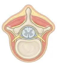
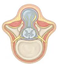
Spina Bifida (Spinal Dysraphism) — Overview
Spina Bifida (Spinal Dysraphism) is a congenital neural tube closure defect occurring between Days 21 and 28 of embryonic gestation (strongly reduced by periconceptional folic acid supplementation), resulting in incomplete fusion of the posterior vertebral arches (most commonly lumbosacral). It encompasses three clinical forms: 1) Spina Bifida Occulta (closed skin, bony cleft only, often tuft of hair/dimple), 2) Meningocele (herniation of meninges/CSF without neural tissue), and 3) Myelomeningocele (open herniation of spinal cord and nerve roots). Rehabilitation is guided by the functional neurological motor level (Thoracic, High Lumbar L1–L2, Mid-Lumbar L3, Low Lumbar L4–L5, or Sacral S1–S2), insensate skin protection, latex-free environment, and Tethered Cord / Shunt surveillance!
Spina bifida (split spine) is a birth condition in which one or more spinal bones in the lower back do not close completely during the first month of pregnancy. Depending on the exact spinal level (especially whether the L3 front-thigh muscle and L4 shin muscle are active), children with spina bifida can achieve wonderful mobility using customized braces, strong upper bodies, and play-based physiotherapy!
Primary Causes & Biomechanics
Key Points You Should Know
- •Failure of caudal neuropore closure during the 4th week of gestation (genetic + folate deficiency factors)
- •Flaccid Lower Motor Neuron Paralysis at and below the myelodysplastic level (sometimes mixed with upper motor neuron signs), producing muscle imbalance-driven orthopedic deformities (hip dislocation, knee flexion/extension contractures, clubfoot, or calcaneovalgus foot)
- •70% Latex Allergy Prevalence & Neurogenic Bladder/Bowel: Repeated early mucosal/surgical exposure sensitizes children to natural rubber latex!
- •Rule #1 — A 100% Latex-Free Environment (Even Exercise Bands & Balloons!): Children with spina bifida have a very high risk of developing a severe latex allergy. Never use natural rubber balloons, latex therapy bands (TheraBand), or latex gloves—always use latex-free synthetic bands and vinyl/foil balloons!
- •Rule #2 — Twice-Daily Skin Checks Inside Shoes & Braces: Because skin feeling is reduced in the legs, Feet, and bottom, a tight brace or rough surface can cause a painless sore. Check the feet, heels, ankles, and bottom every morning and evening!
- •Tummy Time Prevents Tight Hips: Spending 20–30 minutes daily playing on the tummy stretches the hips straight so standing and walking braces fit comfortably.
Assessment Methods
1. Goniometric ROM & Postural / Kinematic Analysis
Functional Neurological Motor Level Assessment: L1–L2: Iliopsoas hip flexion; L3: Quadriceps knee extension (key threshold for household/community walking with AFOs/crutches!); L4: Tibialis anterior ankle dorsiflexion/inversion; L5: Extensor hallucis longus & gluteus medius; S1–S2: Gastrocnemius-soleus plantarflexion.
2. Special Clinical Provocation & Neurological/Functional Tests
Hip stability (Ortolani/Barlow/abduction), knee/ankle contracture goniometry, sitting trunk balance, and daily insensate skin inspection.
3. Standardized Clinical Outcome Scales & Imaging Criteria
Pediatric Evaluation of Disability Inventory (PEDI), Hoffer Functional Ambulation Scale, and Spine MRI / Renal-Bladder Ultrasound.
1. Range of Motion & Posture Check
Your physiotherapist gently evaluates how comfortably and smoothly your embryonic neural tube (vertebral arch closure at Days 21–28 of gestation), spinal cord/cauda equina, and lower extremity myotomes moves and checks your overall posture.
2. Muscle Strength & Functional Movement Check
Your muscle balance, walking or reaching comfort, and specific gentle movement tests are evaluated without triggering sharp pain.
3. Typical Symptoms & Daily Impact Review
Flaccid (Loose) Weakness & Sensory Loss Below the Lesion Level: Varying from full paralysis of the legs (high lumbar/thoracic) to isolated weakness in tiptoe push-off and outer hip balance (low lumbar L4–S1).
Treatment Options & Phase-Based Protocol
✨ Enriched GIF Exercise & Precaution GuidePhase 1: Acute Symptom Modulation, Tissue Protection & Early Activation
- •Phase 1 Clinical Focus & Tissue Protection: Phase 1 — Insensate Skin Care, Latex-Free Contracture Prevention & Prone Phase: Use 100% LATEX-FREE gloves, bands, mats, and toys! Perform daily gentle passive/active-assisted hip, knee, and ankle ROM (ankle pumps, towel stretches, supine heel slides) and daily prone tummy-time to prevent hip/knee flexion contractures.
- •Pain, Edema & Neuromuscular Modulation: Targeted physical modalities, relative load management, and early protected mobility of the embryonic neural tube (vertebral arch closure at Days 21–28 of gestation), spinal cord/cauda equina, and lower extremity myotomes without provoking mechanical or inflammatory irritation.
- •Proximal & Distal Kinetic Chain Maintenance: Maintain active mobility and circulation in adjacent joints while protecting the primary healing structures in Spina Bifida.
Phase 2: Progressive Mobility, Motor Control & Targeted Strengthening
- •Phase 2 Progressive Mobilization & Motor Control: Phase 2 — Upper Extremity Hypertrophy, Trunk Control & Transfer Phase: Seated push-ups (for transfer independence and every-20-min pressure relief!), Bobath bilateral overhead reaching & quadruped/seated core stabilization, and thoracic expansion breathing (preventing neuromuscular scoliosis).
- •Biomechanical Alignment & Co-Contraction: Restore balanced agonist-antagonist force couples, joint centration, and multi-planar neuromuscular control across the embryonic neural tube (vertebral arch closure at Days 21–28 of gestation), spinal cord/cauda equina, and lower extremity myotomes.
- •Myofascial Extensibility & Endurance: Address secondary compensatory tightness and build local muscular endurance within safe, symptom-free biomechanical parameters.
Phase 3: Functional Integration, Proprioception & Long-Term Prevention
- •Phase 3 Functional Integration & Long-Term Prevention: Phase 3 — Level-Matched Orthotic Standing & Ambulation Phase: Thoracic/L1–L2: Standing frame / RGO / HKAFO + wheelchair skills; L3: KAFO -> AFO + crutches; L4–L5: Solid/Floor-Reaction AFO (compensating for absent S1 calf push-off and preventing crouch gait!); S1–S2: SMO/foot orthoses.
- •Proprioceptive Balance & Dynamic Loading: Progress to task-specific closed-chain, balance, and functional movement patterns tailored to daily life, occupational, or athletic demands.
- •Self-Management & Relapse Prophylaxis: Establish lifelong ergonomic habits, home exercise maintenance, and early warning-sign awareness to prevent recurrence.
Step 1: Calming Pain, Protecting the Area & Gentle Starter Motion
- •Rule #1 — A 100% Latex-Free Environment (Even Exercise Bands & Balloons!): Children with spina bifida have a very high risk of developing a severe latex allergy. Never use natural rubber balloons, latex therapy bands (TheraBand), or latex gloves—always use latex-free synthetic bands and vinyl/foil balloons!
- •Calm Protection & Gentle Start: During the initial stage of Split Spine / Spina Bifida, avoid painful overloading and perform the gentle, pain-free starter movements shown below to support circulation and comfort.
- •Respecting Your Body's Signals: Every movement should stay within a comfortable, non-painful zone—never force through sharp or radiating pain.
Step 2: Restoring Flexibility & Building Supporting Muscle Strength
- •Rule #2 — Twice-Daily Skin Checks Inside Shoes & Braces: Because skin feeling is reduced in the legs, Feet, and bottom, a tight brace or rough surface can cause a painless sore. Check the feet, heels, ankles, and bottom every morning and evening!
- •Step-by-Step Flexibility & Support: As acute sensitivity settles, gradually build mobility and strengthen the supporting muscles around the joint at a steady, controlled pace.
- •Posture & Breathing Coordination: Keep your posture upright and breathe smoothly (never hold your breath!) during every repetition.
Step 3: Safe Daily Habits, Stamina & Preventing Recurrence
- •Tummy Time Prevents Tight Hips: Spending 20–30 minutes daily playing on the tummy stretches the hips straight so standing and walking braces fit comfortably.
- •Confident Return to Daily Life: Integrate your stronger muscles and improved balance into walking, stairs, work, and recreational activities.
- •Preventing Future Flare-Ups: Continue your core protective habits and key maintenance exercises even after symptoms resolve.
Animated (GIF) Therapeutic Exercise Guide
Animated clinical exercise protocol with biomechanical angles, target muscle groups, and dosage parameters (Click image to enlarge)
Step-by-step animated exercise demonstrations you can safely perform at home (Click image to enlarge)
 🔍 Enlarge
1. Supine Active-Assisted Heel Slide & Synovial Diffusion Mobilization
1. Supine Heel Slide (Gentle Knee & Hip Bending)
Controlled flexion and extension of the lower extremity by sliding the heel along the support surface without axial weight-bearing. Stimulates synovial fluid diffusion across avascular cartilage and prevents capsular adhesions. Specifically indicated in Spina Bifida rehabilitation to optimize embryonic neural tube (vertebral arch closure at Days 21–28 of gestation), spinal cord/cauda equina, and lower extremity myotomes biomechanics and neuromuscular control.
While lying on your back, slowly slide your heel along the bed sheet toward your hip within a comfortable, pain-free angle, hold for 3 seconds, and gently slide it back out straight. Perform smoothly within a comfortable, pain-free range to support your Split Spine / Spina Bifida recovery.
 🔍 Enlarge
2. Rhythmic Active Ankle Dorsiflexion & Plantarflexion Pumping (Venous/Edema Pump)
2. Ankle Toe-Pulls and Pointing (Circulation & Swelling Pump)
Rhythmic alternating contraction of the tibialis anterior and gastrocnemius-soleus complex. Activates the deep venous calf pump to prevent deep vein thrombosis (DVT), clears distal edema, and maintains talocrural mobility. Specifically indicated in Spina Bifida rehabilitation to optimize embryonic neural tube (vertebral arch closure at Days 21–28 of gestation), spinal cord/cauda equina, and lower extremity myotomes biomechanics and neuromuscular control.
While lying down or sitting with your legs supported, pull your toes and feet firmly upward toward your shins, hold for 3 seconds, then point them downward like pressing a gas pedal. Perform smoothly within a comfortable, pain-free range to support your Split Spine / Spina Bifida recovery.
 🔍 Enlarge
3. Scapulothoracic Retraction & Depression (Middle/Lower Trapezius & Rhomboids)
3. Shoulder Blade Squeeze (Postural Alignment)
Adduction and depression of the scapulae via the middle/lower trapezius and rhomboids while inhibiting upper trapezius overactivity. Corrects anterior scapular tilt, widens the subacromial outlet, and unloads the anterior thoracic column. Specifically indicated in Spina Bifida rehabilitation to optimize embryonic neural tube (vertebral arch closure at Days 21–28 of gestation), spinal cord/cauda equina, and lower extremity myotomes biomechanics and neuromuscular control.
Sit or stand tall. Keep your shoulders relaxed away from your ears, gently draw your shoulder blades together and slightly downward toward your back pockets, hold for 5 seconds, and release. Perform smoothly within a comfortable, pain-free range to support your Split Spine / Spina Bifida recovery.
 🔍 Enlarge
4. Prone Positioning & Controlled Thoracolumbar Extension (McKenzie / Back Extensor)
4. Prone Lying & Gentle Back Extension Lift
Prone positioning and graduated thoracic/lumbar extension (with a pillow under the abdomen when indicated for spinal unloading). Counters flexed postural kyphosis, stretches tight hip flexors, and strengthens paraspinal extensors. Specifically indicated in Spina Bifida rehabilitation to optimize embryonic neural tube (vertebral arch closure at Days 21–28 of gestation), spinal cord/cauda equina, and lower extremity myotomes biomechanics and neuromuscular control.
Lie on your stomach (place a small pillow under your hips/tummy if needed for comfort). Gently lift your head and chest slightly off the mat by squeezing your back muscles without straining your neck, hold for 3–5 seconds, and lower. Perform smoothly within a comfortable, pain-free range to support your Split Spine / Spina Bifida recovery.
 🔍 Enlarge
5. Seated Depression Transfer Lift ('Push-Up' — Triceps, Latissimus & Pressure Relief)
5. Seated Arm-Press Trunk Lift (Transfer & Pressure Relief)
Pressing down on armrests or push-blocks with elbows extending and scapulae depressing to lift the buttocks clear of the seat. Essential for independent bed-to-wheelchair transfers, crutch ambulation, and relieving ischial tuberosity pressure every 20–30 minutes. Specifically indicated in Spina Bifida rehabilitation to optimize embryonic neural tube (vertebral arch closure at Days 21–28 of gestation), spinal cord/cauda equina, and lower extremity myotomes biomechanics and neuromuscular control.
While sitting securely in your chair or wheelchair with brakes locked, place your hands on the armrests and push down to lift your hips slightly off the seat for 5–10 seconds, then lower gently. Perform smoothly within a comfortable, pain-free range to support your Split Spine / Spina Bifida recovery.
 🔍 Enlarge
6. Diaphragmatic & Pursed-Lip Breathing (PLB — Autonomic & Ventilatory Control)
6. Calm Belly Breathing with Pursed-Lip Exhale
Slow nasal inhalation expanding the lower ribcage and abdomen (while relaxing accessory neck/shoulder muscles), followed by prolonged expiration through pursed lips at a 1:2 ratio. Prevents dynamic airway collapse, improves basal ventilation, and reduces sympathetic tone. Specifically indicated in Spina Bifida rehabilitation to optimize embryonic neural tube (vertebral arch closure at Days 21–28 of gestation), spinal cord/cauda equina, and lower extremity myotomes biomechanics and neuromuscular control.
Sit comfortably and place one hand on your chest and the other on your belly. Breathe in slowly through your nose for 2–3 seconds so your belly gently rises, then breathe out slowly for 4–6 seconds through pursed lips as if flickering a candle. Perform smoothly within a comfortable, pain-free range to support your Split Spine / Spina Bifida recovery.
Movements to Avoid & Strict Precautions
1. Using Natural Rubber Latex Balloons, Latex Exercise Bands, or Latex Toys (Severe Anaphylaxis Risk!)
1. Allowing the child to play with rubber balloons or using standard latex exercise bands (ALWAYS use 100% latex-free equipment!)
2. Crawling or Scooting on Bare Floors Without Pants/Knee Pads (Scrapes Insensate Skin!)
2. Letting the child scoot on rough carpets or hot tiles with bare legs/feet
3. Putting On AFO/KAFO Braces Without Checking Skin for Red Pressure Marks
3. Putting braces on without checking the heels and ankles for red spots
4. Forcefully Stretching Osteopenic Insensate Legs (High Risk of Painless Femur/Tibia Fracture!)
4. Forcefully pushing on stiff legs that lack pain sensation
Spina Bifida (Spinal Dysraphism) — Video Guide
Click directly on any video to open and watch in a window, or review details via 📋 Summary & Options.
Condition Overview Video (1 Video)
Summary of pathophysiology, biomechanics, and clinical presentation
What the condition is, why it develops, and its basic mechanism

What Are Spina Bifida and Myelomeningocele? Clinical Features
Spina Bifida (Spinal Dysraphism) is a congenital neural tube closure defect occurring between Days 21 and 28 of embryonic gestation (strongly reduced by periconceptional folic acid supplementation), resulting in incomplete fusion of the posterior vertebral arches (most commonly lumbosacral). It encompasses three clinical forms: 1) Spina Bifida Occulta (closed skin, bony cleft only, often tuft of hair/dimple), 2) Meningocele (herniation of meninges/CSF without neural tissue), and 3) Myelomeningocele (open herniation of spinal cord and nerve roots). Rehabilitation is guided by the functional neurological motor level (Thoracic, High Lumbar L1–L2, Mid-Lumbar L3, Low Lumbar L4–L5, or Sacral S1–S2), insensate skin protection, latex-free environment, and Tethered Cord / Shunt surveillance!
Spina bifida (split spine) is a birth condition in which one or more spinal bones in the lower back do not close completely during the first month of pregnancy. Depending on the exact spinal level (especially whether the L3 front-thigh muscle and L4 shin muscle are active), children with spina bifida can achieve wonderful mobility using customized braces, strong upper bodies, and play-based physiotherapy!
Treatment and Exercise Videos
Phase-based physiotherapy protocols, mobilization, and therapeutic exercises
Step-by-step recovery and safe home exercise applications

Spina Bifida and Myelomeningocele Rehabilitation: Orthotics and Gait Training
Phase 1: Acute Symptom Modulation, Tissue Protection & Early Activation: Phase 1 Clinical Focus & Tissue Protection: Phase 1 — Insensate Skin Care, Latex-Free Contracture Prevention & Prone Phase: Use 100% LATEX-FREE gloves, bands, mats, and toys! Perform daily gentle passive/active-assisted hip, knee, and ankle ROM (ankle pumps, towel stretches, supine heel slides) and daily prone tummy-time to prevent hip/knee flexion contractures.
Step 1: Calming Pain, Protecting the Area & Gentle Starter Motion: Rule #1 — A 100% Latex-Free Environment (Even Exercise Bands & Balloons!): Children with spina bifida have a very high risk of developing a severe latex allergy. Never use natural rubber balloons, latex therapy bands (TheraBand), or latex gloves—always use latex-free synthetic bands and vinyl/foil balloons!

Home Exercises in Pediatric Neurological Conditions (CP, Spina Bifida, Brachial Plexus)
Phase 2: Progressive Mobility, Motor Control & Targeted Strengthening: Phase 2 Progressive Mobilization & Motor Control: Phase 2 — Upper Extremity Hypertrophy, Trunk Control & Transfer Phase: Seated push-ups (for transfer independence and every-20-min pressure relief!), Bobath bilateral overhead reaching & quadruped/seated core stabilization, and thoracic expansion breathing (preventing neuromuscular scoliosis).
Step 2: Restoring Flexibility & Building Supporting Muscle Strength: Rule #2 — Twice-Daily Skin Checks Inside Shoes & Braces: Because skin feeling is reduced in the legs, Feet, and bottom, a tight brace or rough surface can cause a painless sore. Check the feet, heels, ankles, and bottom every morning and evening!
Clinical Assessment, Goniometry & Special Test Videos
Displayed only in Healthcare Professional mode (hidden in Patient mode)
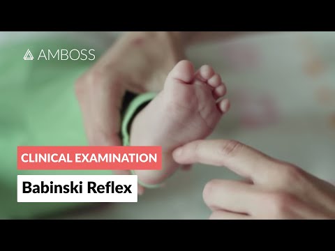
Pediatric Neurological Examination: Babinski and Plantar Reflex Assessment
Goniometric ROM & Postural / Kinematic Analysis: Functional Neurological Motor Level Assessment: L1–L2: Iliopsoas hip flexion; L3: Quadriceps knee extension (key threshold for household/community walking with AFOs/crutches!); L4: Tibialis anterior ankle dorsiflexion/inversion; L5: Extensor hallucis longus & gluteus medius; S1–S2: Gastrocnemius-soleus plantarflexion.

Manual Muscle Testing (MRC 0–5 Grading) — Clinical Motor Strength Examination
Special Clinical Provocation & Neurological/Functional Tests: Hip stability (Ortolani/Barlow/abduction), knee/ankle contracture goniometry, sitting trunk balance, and daily insensate skin inspection.

Hip Joint Goniometric Measurement (Flexion, Extension, Abduction, Rotations)
Standardized Clinical Outcome Scales & Imaging Criteria: Pediatric Evaluation of Disability Inventory (PEDI), Hoffer Functional Ambulation Scale, and Spine MRI / Renal-Bladder Ultrasound.