Osteogenesis Imperfecta (Brittle Bone Disease)Brittle Bone Disease (Osteogenesis Imperfecta)
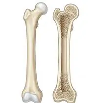
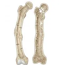
Osteogenesis Imperfecta (Brittle Bone Disease) — Overview
Osteogenesis Imperfecta (OI / 'Brittle Bone Disease') is a hereditary connective tissue disorder caused (in ~85–90% of cases) by autosomal dominant mutations in the COL1A1 or COL1A2 genes encoding Type I procollagen—causing either a quantitative defect (50% normal collagen -> Sillence Type I Mild OI with distinct Blue Sclerae) or a qualitative structural defect (abnormal triple-helix glycine substitution -> Type III Severe Progressive Deformity OI or Type IV Moderate OI). THE PARADOX & GOLDEN LAW OF OI REHABILITATION: Immobilization and fear of movement cause rapid disuse osteoporosis, which triggers EVEN MORE fractures (the fracture-immobilization-osteopenia vicious cycle)! Conversely, safe active muscle pull, hydrotherapy, and vertical axial loading (with Fassier-Duval telescopic intramedullary rods + bisphosphonate therapy) build cortical bone thickness and PREVENT fractures!
Osteogenesis Imperfecta (OI / 'brittle bone disease') is a genetic condition affecting Type I collagen—the protein mesh that gives bones their flexible strength. The most important discovery in modern OI care is that wrapping a child in cotton wool without movement actually makes bones thinner and more fragile! Safe swimming/warm-pool play, active muscle strengthening, and supported upright standing (alongside medical bone-strengthening treatments) act like an internal shield that makes bones thicker and prevents fractures!
Primary Causes & Biomechanics
Key Points You Should Know
- •Defective or deficient Type I collagen in bone osteoid matrix, ligaments (causing generalized joint hypermobility and pes planus), sclerae (blue sclerae), and teeth (dentinogenesis imperfecta)
- •Mechanical Rule for Handling & Exercise (Never Use Long Lever Arms or Torsion!): Bone is strongest against axial compression (standing upright) and weakest against torsional twisting and long-lever bending! Therefore, resistance or handling must ALWAYS be applied PROXIMALLY (close to the hip/shoulder joint), NEVER at the ankle or wrist!
- •Multidisciplinary standard of care: cyclic IV Bisphosphonates (Pamidronate/Zoledronate) + telescopic intramedullary rodding + active hydrotherapy & weight-bearing physiotherapy
- •Golden Handling Rule for Babies — 'Scoop the Bottom, Never Pull the Ankles!': When changing a diaper, NEVER lift your baby by the ankles—instead, gently roll your baby onto their side or slide your whole flat hand under their buttocks! When picking them up, spread your fingers wide under their back, bottom, and head (never lift under the armpits).
- •Why Warm Water (Hydrotherapy) Is Magical in OI: In a warm pool, water cushions every movement and removes the risk of falling, while gently resisting your child's kicks and arm strokes so their muscles pull on the bones and make them stronger!
- •Keep Cast Time as Short as Safely Possible: After a fracture, sitting immobile for months drains calcium out of the bones. Ask your OI specialist about lightweight splints and starting pool/active movement as soon as early callus forms.
Assessment Methods
1. Goniometric ROM & Postural / Kinematic Analysis
Gross Motor Function Measure (GMFM) / BAMF (Brief Assessment of Motor Function), active ROM (never force passive end-range!), and spine/long-bone bowing assessment.
2. Special Clinical Provocation & Neurological/Functional Tests
Beighton Hypermobility Score, DEXA Lumbar Spine Z-score, and functional ambulation / transfer independence.
3. Standardized Clinical Outcome Scales & Imaging Criteria
Sillence Classification (Types I–IV), DEXA Z-score, and radiographic long-bone bowing / vertebral reshaping.
1. Range of Motion & Posture Check
Your physiotherapist gently evaluates how comfortably and smoothly your Type I collagen matrix (COL1A1 / COL1A2 genes) in cortical/trabecular bone, sclerae, ligaments, dentin, and inner ear moves and checks your overall posture.
2. Muscle Strength & Functional Movement Check
Your muscle balance, walking or reaching comfort, and specific gentle movement tests are evaluated without triggering sharp pain.
3. Typical Symptoms & Daily Impact Review
Recurrent Fractures from Minor Trauma (or even diaper changes/rolling in severe Type III), accompanied by blue-gray tint to the whites of the eyes (blue sclerae).
Treatment Options & Phase-Based Protocol
✨ Enriched GIF Exercise & Precaution GuidePhase 1: Acute Symptom Modulation, Tissue Protection & Early Activation
- •Phase 1 Clinical Focus & Tissue Protection: Phase 1 — Safe Infant Handling ('Scoop, Never Pull!') & Aquatic Hydrotherapy Phase: Teach parents safe handling: NEVER lift an infant under the armpits or pull by the ankles during diaper changes (roll the baby onto the side or scoop under the whole pelvis and shoulders on a padded pillow!); initiate warm-water Hydrotherapy (water supports fragile limbs while providing gentle 360° muscular resistance!) and active unloaded kicking/reaching.
- •Pain, Edema & Neuromuscular Modulation: Targeted physical modalities, relative load management, and early protected mobility of the Type I collagen matrix (COL1A1 / COL1A2 genes) in cortical/trabecular bone, sclerae, ligaments, dentin, and inner ear without provoking mechanical or inflammatory irritation.
- •Proximal & Distal Kinetic Chain Maintenance: Maintain active mobility and circulation in adjacent joints while protecting the primary healing structures in Osteogenesis Imperfecta.
Phase 2: Progressive Mobility, Motor Control & Targeted Strengthening
- •Phase 2 Progressive Mobilization & Motor Control: Phase 2 — Active Muscle Strengthening (Wolff's Law) & Core Phase: Let the child move ACTIVELY against gravity (muscle contraction pulls on periosteal insertions to thicken bone cortex!): supine pelvic bridging, active heel slides, quadruped/tummy-time head & back extension (preventing vertebral compression fractures!), and diaphragmatic/costal breathing.
- •Biomechanical Alignment & Co-Contraction: Restore balanced agonist-antagonist force couples, joint centration, and multi-planar neuromuscular control across the Type I collagen matrix (COL1A1 / COL1A2 genes) in cortical/trabecular bone, sclerae, ligaments, dentin, and inner ear.
- •Myofascial Extensibility & Endurance: Address secondary compensatory tightness and build local muscular endurance within safe, symptom-free biomechanical parameters.
Phase 3: Functional Integration, Proprioception & Long-Term Prevention
- •Phase 3 Functional Integration & Long-Term Prevention: Phase 3 — Graduated Vertical Axial Weight-Bearing (Standing Frame / Rodded Ambulation) Phase: Supported upright standing (tilt-table / standing frame / lightweight HKAFO-AFOs) to load the femurs and tibias along their vertical mechanical axis, controlled chair sit-to-stand, and early mobilization after fractures (minimizing cast time so bone density is not lost!).
- •Proprioceptive Balance & Dynamic Loading: Progress to task-specific closed-chain, balance, and functional movement patterns tailored to daily life, occupational, or athletic demands.
- •Self-Management & Relapse Prophylaxis: Establish lifelong ergonomic habits, home exercise maintenance, and early warning-sign awareness to prevent recurrence.
Step 1: Calming Pain, Protecting the Area & Gentle Starter Motion
- •Golden Handling Rule for Babies — 'Scoop the Bottom, Never Pull the Ankles!': When changing a diaper, NEVER lift your baby by the ankles—instead, gently roll your baby onto their side or slide your whole flat hand under their buttocks! When picking them up, spread your fingers wide under their back, bottom, and head (never lift under the armpits).
- •Calm Protection & Gentle Start: During the initial stage of Brittle Bone Disease, avoid painful overloading and perform the gentle, pain-free starter movements shown below to support circulation and comfort.
- •Respecting Your Body's Signals: Every movement should stay within a comfortable, non-painful zone—never force through sharp or radiating pain.
Step 2: Restoring Flexibility & Building Supporting Muscle Strength
- •Why Warm Water (Hydrotherapy) Is Magical in OI: In a warm pool, water cushions every movement and removes the risk of falling, while gently resisting your child's kicks and arm strokes so their muscles pull on the bones and make them stronger!
- •Step-by-Step Flexibility & Support: As acute sensitivity settles, gradually build mobility and strengthen the supporting muscles around the joint at a steady, controlled pace.
- •Posture & Breathing Coordination: Keep your posture upright and breathe smoothly (never hold your breath!) during every repetition.
Step 3: Safe Daily Habits, Stamina & Preventing Recurrence
- •Keep Cast Time as Short as Safely Possible: After a fracture, sitting immobile for months drains calcium out of the bones. Ask your OI specialist about lightweight splints and starting pool/active movement as soon as early callus forms.
- •Confident Return to Daily Life: Integrate your stronger muscles and improved balance into walking, stairs, work, and recreational activities.
- •Preventing Future Flare-Ups: Continue your core protective habits and key maintenance exercises even after symptoms resolve.
Animated (GIF) Therapeutic Exercise Guide
Animated clinical exercise protocol with biomechanical angles, target muscle groups, and dosage parameters (Click image to enlarge)
Step-by-step animated exercise demonstrations you can safely perform at home (Click image to enlarge)
 🔍 Enlarge
1. Supine Active-Assisted Heel Slide & Synovial Diffusion Mobilization
1. Supine Heel Slide (Gentle Knee & Hip Bending)
Controlled flexion and extension of the lower extremity by sliding the heel along the support surface without axial weight-bearing. Stimulates synovial fluid diffusion across avascular cartilage and prevents capsular adhesions. Specifically indicated in Osteogenesis Imperfecta rehabilitation to optimize Type I collagen matrix (COL1A1 / COL1A2 genes) in cortical/trabecular bone, sclerae, ligaments, dentin, and inner ear biomechanics and neuromuscular control.
While lying on your back, slowly slide your heel along the bed sheet toward your hip within a comfortable, pain-free angle, hold for 3 seconds, and gently slide it back out straight. Perform smoothly within a comfortable, pain-free range to support your Brittle Bone Disease recovery.
 🔍 Enlarge
2. Isometric Quadriceps Setting (Popliteal Towel Press)
2. Under-Knee Towel Press (Quadriceps Tightening)
Isometric compression of a rolled towel beneath the popliteal fossa into the surface with simultaneous ankle dorsiflexion. Activates the Vastus Medialis Obliquus (VMO) and overcomes arthrogenic muscle inhibition with 0° joint shear stress. Specifically indicated in Osteogenesis Imperfecta rehabilitation to optimize Type I collagen matrix (COL1A1 / COL1A2 genes) in cortical/trabecular bone, sclerae, ligaments, dentin, and inner ear biomechanics and neuromuscular control.
Place a small rolled towel under your knee while lying on your back or sitting with your leg straight. Pull your toes toward you while firmly pressing the back of your knee down into the towel, hold for 5 seconds, and relax while exhaling. Perform smoothly within a comfortable, pain-free range to support your Brittle Bone Disease recovery.
 🔍 Enlarge
3. Scapulothoracic Retraction & Depression (Middle/Lower Trapezius & Rhomboids)
3. Shoulder Blade Squeeze (Postural Alignment)
Adduction and depression of the scapulae via the middle/lower trapezius and rhomboids while inhibiting upper trapezius overactivity. Corrects anterior scapular tilt, widens the subacromial outlet, and unloads the anterior thoracic column. Specifically indicated in Osteogenesis Imperfecta rehabilitation to optimize Type I collagen matrix (COL1A1 / COL1A2 genes) in cortical/trabecular bone, sclerae, ligaments, dentin, and inner ear biomechanics and neuromuscular control.
Sit or stand tall. Keep your shoulders relaxed away from your ears, gently draw your shoulder blades together and slightly downward toward your back pockets, hold for 5 seconds, and release. Perform smoothly within a comfortable, pain-free range to support your Brittle Bone Disease recovery.
 🔍 Enlarge
4. Functional Closed-Kinetic-Chain Chair Transfer (Sit-to-Stand Biomechanics)
4. Controlled Chair Sit-to-Stand Transfer
Controlled transition from sitting to standing and eccentric lowering back to the chair with symmetrical weight distribution, anterior trunk inclination ('nose over toes'), and neutral frontal knee alignment. Specifically indicated in Osteogenesis Imperfecta rehabilitation to optimize Type I collagen matrix (COL1A1 / COL1A2 genes) in cortical/trabecular bone, sclerae, ligaments, dentin, and inner ear biomechanics and neuromuscular control.
Sit near the front of a sturdy chair with your feet hip-width apart. Lean slightly forward and push evenly through both heels to stand up tall, then slowly lower yourself back down over 3 seconds. Perform smoothly within a comfortable, pain-free range to support your Brittle Bone Disease recovery.
 🔍 Enlarge
5. Straight Leg Raise (SLR — Multi-Plane Isometric Knee Lock)
5. Supine Straight Leg Raise
Maintaining the knee locked in full extension (0°) with ankle dorsiflexion while elevating the lower extremity to 45° hip flexion. Strengthens the quadriceps and iliopsoas complex without compressing or shearing the articular surfaces. Specifically indicated in Osteogenesis Imperfecta rehabilitation to optimize Type I collagen matrix (COL1A1 / COL1A2 genes) in cortical/trabecular bone, sclerae, ligaments, dentin, and inner ear biomechanics and neuromuscular control.
Bend your healthy knee with the foot flat on the bed. Tighten the thigh muscle of your affected leg to lock the knee straight, lift the leg to the height of the opposite knee, hold for 3 seconds, and lower slowly. Perform smoothly within a comfortable, pain-free range to support your Brittle Bone Disease recovery.
 🔍 Enlarge
6. Diaphragmatic & Pursed-Lip Breathing (PLB — Autonomic & Ventilatory Control)
6. Calm Belly Breathing with Pursed-Lip Exhale
Slow nasal inhalation expanding the lower ribcage and abdomen (while relaxing accessory neck/shoulder muscles), followed by prolonged expiration through pursed lips at a 1:2 ratio. Prevents dynamic airway collapse, improves basal ventilation, and reduces sympathetic tone. Specifically indicated in Osteogenesis Imperfecta rehabilitation to optimize Type I collagen matrix (COL1A1 / COL1A2 genes) in cortical/trabecular bone, sclerae, ligaments, dentin, and inner ear biomechanics and neuromuscular control.
Sit comfortably and place one hand on your chest and the other on your belly. Breathe in slowly through your nose for 2–3 seconds so your belly gently rises, then breathe out slowly for 4–6 seconds through pursed lips as if flickering a candle. Perform smoothly within a comfortable, pain-free range to support your Brittle Bone Disease recovery.
Movements to Avoid & Strict Precautions
1. Lifting an Infant by the Ankles (During Diaper Changes) or Under the Armpits (Causes Rib and Femur Fractures!)
1. NEVER pull a baby by the ankles to change a diaper or lift them under the armpits (always scoop under the bottom and torso!)
2. Applying Manual Resistance at the Ankle or Wrist (Long Lever Arm Breaks the Bone Shaft!)
2. Pushing or pulling against the child's hand or ankle (never force passive stretches or apply long-lever resistance!)
3. Twisting (Rotational Torsion) or Spinal Flexion (Sit-Ups / Forward Bending)
3. Twisting the arms/legs, doing sit-ups, or jumping on trampolines/hard floors
4. Prolonged Bed Rest After Fractures (Accelerates Bone Loss and Causes a 'Fracture Cycle'!)
4. Keeping the child completely immobile in bed for months after a fracture heals
Osteogenesis Imperfecta (Brittle Bone Disease) — Video Guide
Click directly on any video to open and watch in a window, or review details via 📋 Summary & Options.
Condition Overview Video (1 Video)
Summary of pathophysiology, biomechanics, and clinical presentation
What the condition is, why it develops, and its basic mechanism

What Is Osteogenesis Imperfecta (Brittle Bone Disease)? Types and Features
Osteogenesis Imperfecta (OI / 'Brittle Bone Disease') is a hereditary connective tissue disorder caused (in ~85–90% of cases) by autosomal dominant mutations in the COL1A1 or COL1A2 genes encoding Type I procollagen—causing either a quantitative defect (50% normal collagen -> Sillence Type I Mild OI with distinct Blue Sclerae) or a qualitative structural defect (abnormal triple-helix glycine substitution -> Type III Severe Progressive Deformity OI or Type IV Moderate OI). THE PARADOX & GOLDEN LAW OF OI REHABILITATION: Immobilization and fear of movement cause rapid disuse osteoporosis, which triggers EVEN MORE fractures (the fracture-immobilization-osteopenia vicious cycle)! Conversely, safe active muscle pull, hydrotherapy, and vertical axial loading (with Fassier-Duval telescopic intramedullary rods + bisphosphonate therapy) build cortical bone thickness and PREVENT fractures!
Osteogenesis Imperfecta (OI / 'brittle bone disease') is a genetic condition affecting Type I collagen—the protein mesh that gives bones their flexible strength. The most important discovery in modern OI care is that wrapping a child in cotton wool without movement actually makes bones thinner and more fragile! Safe swimming/warm-pool play, active muscle strengthening, and supported upright standing (alongside medical bone-strengthening treatments) act like an internal shield that makes bones thicker and prevents fractures!
Treatment and Exercise Videos
Phase-based physiotherapy protocols, mobilization, and therapeutic exercises
Step-by-step recovery and safe home exercise applications

Safe Physiotherapy and Fracture Prevention in Osteogenesis Imperfecta
Phase 1: Acute Symptom Modulation, Tissue Protection & Early Activation: Phase 1 Clinical Focus & Tissue Protection: Phase 1 — Safe Infant Handling ('Scoop, Never Pull!') & Aquatic Hydrotherapy Phase: Teach parents safe handling: NEVER lift an infant under the armpits or pull by the ankles during diaper changes (roll the baby onto the side or scoop under the whole pelvis and shoulders on a padded pillow!); initiate warm-water Hydrotherapy (water supports fragile limbs while providing gentle 360° muscular resistance!) and active unloaded kicking/reaching.
Step 1: Calming Pain, Protecting the Area & Gentle Starter Motion: Golden Handling Rule for Babies — 'Scoop the Bottom, Never Pull the Ankles!': When changing a diaper, NEVER lift your baby by the ankles—instead, gently roll your baby onto their side or slide your whole flat hand under their buttocks! When picking them up, spread your fingers wide under their back, bottom, and head (never lift under the armpits).

Trunk Control, Balance, and Functional Home Exercises in Pediatric Rehabilitation
Phase 2: Progressive Mobility, Motor Control & Targeted Strengthening: Phase 2 Progressive Mobilization & Motor Control: Phase 2 — Active Muscle Strengthening (Wolff's Law) & Core Phase: Let the child move ACTIVELY against gravity (muscle contraction pulls on periosteal insertions to thicken bone cortex!): supine pelvic bridging, active heel slides, quadruped/tummy-time head & back extension (preventing vertebral compression fractures!), and diaphragmatic/costal breathing.
Step 2: Restoring Flexibility & Building Supporting Muscle Strength: Why Warm Water (Hydrotherapy) Is Magical in OI: In a warm pool, water cushions every movement and removes the risk of falling, while gently resisting your child's kicks and arm strokes so their muscles pull on the bones and make them stronger!
Clinical Assessment, Goniometry & Special Test Videos
Displayed only in Healthcare Professional mode (hidden in Patient mode)

Hip Joint Goniometric Measurement (Flexion, Extension, Abduction, Rotations)
Goniometric ROM & Postural / Kinematic Analysis: Gross Motor Function Measure (GMFM) / BAMF (Brief Assessment of Motor Function), active ROM (never force passive end-range!), and spine/long-bone bowing assessment.

Knee Joint Goniometric Measurement (Active & Passive Flexion / Extension ROM)
Special Clinical Provocation & Neurological/Functional Tests: Beighton Hypermobility Score, DEXA Lumbar Spine Z-score, and functional ambulation / transfer independence.