Developmental Dysplasia of the Hip (Pediatric DDH)Congenital Hip Dislocation (Developmental Dysplasia of the Hip (DDH))
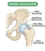
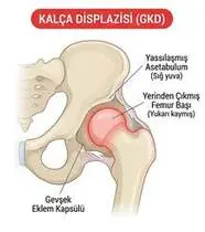
Developmental Dysplasia of the Hip (Pediatric DDH) — Overview
Pediatric Developmental Dysplasia of the Hip (Infantile DDH) spans a spectrum from acetabular dysplasia (shallow, immature cartilaginous socket) and subluxatable hip (Barlow positive) to frank dislocation (Ortolani positive) in newborns and infants. In the first 6 months of life, the acetabulum and femoral head are predominantly cartilaginous and possess extraordinary remodeling potential—maintaining the femoral head concentrically reduced inside the acetabulum in the 'Human Position' (90–100° hip flexion and 45–60° gentle abduction via a Pavlik Harness) stimulates normal deepening of the acetabular roof in >90–95% of cases without surgery! CRITICAL SAFETY RULE: Extreme forced abduction (>60° 'Frog-Leg' cast/splint position) compresses the medial femoral circumflex artery and causes Avascular Necrosis (AVN) of the femoral head!
Pediatric Developmental Dysplasia of the Hip (infantile DDH / congenital hip dislocation) is a condition in newborns and babies where the hip socket is shallow, allowing the ball of the thighbone to slip partially or fully out. When caught in the first 6 months, keeping the baby's hips gently bent and apart in the natural 'M-Position' (with a Pavlik harness and hip-healthy carrying) allows the baby's growing socket to mold into a deep, 100% normal hip!
Primary Causes & Biomechanics
Key Points You Should Know
- •Risk factors ('The 5 F's'): Female sex (80%, due to maternal relaxin sensitivity), Firstborn (tight primigravida uterus), Frank Breech presentation (#1 mechanical risk factor!), Family history, and Oligohydramnios / Post-birth tight straight-leg swaddling!
- •Why Traditional Tight Straight-Leg Swaddling Causes Hip Dislocation: Binding a newborn's legs straight together (hip extension + adduction) uses the iliopsoas and adductors as a lever to pry the femoral head directly out of the shallow socket!
- •After 6 months of age (once the femoral head ossifies): failure of Pavlik harness requires closed reduction + hip spica cast or open reduction / Salter/Pemberton pelvic osteotomy
- •Rule #1 — NEVER Swaddle Your Baby's Legs Straight and Tight! Wrapping a baby's legs straight and pressed together like a cigar forces the hip ball right out of its shallow socket! Always use a hip-healthy sleep sack where the legs have plenty of room to bend up and out like a little frog ('M-Position').
- •The Pavlik Harness Golden Rule (Never Force the Knees Flat!): The Pavlik harness works by letting the baby kick gently inside a safe zone. Never tighten the back straps to force the knees flat against the bed—extreme wide flattening pinches the delicate blood vessels to the hip ball!
- •Checking Thigh Folds & Knee Height: When changing diapers, check that both knees open out to the sides equally and sit at the same height when bent.
Assessment Methods
1. Goniometric ROM & Postural / Kinematic Analysis
Ortolani Maneuver (gently abducting and lifting the greater trochanter reduces a *dislocated* femoral head back into the socket with a palpable 'clunk'—valid in the first 1–3 months!), Barlow Maneuver (gentle posterior pressure on an adducted hip detects an *unstable/dislocatable* hip), and Hip Abduction Symmetry in 90° Flexion (after 2–3 months, unilateral limitation of hip abduction <45–60° is the MOST RELIABLE clinical sign of DDH!).
2. Special Clinical Provocation & Neurological/Functional Tests
Galeazzi (Allis) Sign (apparent femoral shortening—one knee sits lower than the other when hips and knees are flexed in supine!), asymmetric gluteal/thigh skin folds, and Graf Hip Ultrasound (before 4–6 months; AP Pelvis X-ray after 4–6 months).
3. Standardized Clinical Outcome Scales & Imaging Criteria
Graf Ultrasonographic Classification (Type I: alpha angle >60° normal; Type II: dysplastic; Type III–IV: subluxated/dislocated), Acetabular Index on X-ray, and hip abduction goniometry.
1. Range of Motion & Posture Check
Your physiotherapist gently evaluates how comfortably and smoothly your infantile acetabulum (cartilaginous roof and labrum) and proximal femoral head epiphysis (Developmental Dysplasia of the Hip — DDH) moves and checks your overall posture.
2. Muscle Strength & Functional Movement Check
Your muscle balance, walking or reaching comfort, and specific gentle movement tests are evaluated without triggering sharp pain.
3. Typical Symptoms & Daily Impact Review
In Newborns (0–3 Months): Painless 'clunk' felt during diaper changes or doctor exam (Ortolani sign) and extra or uneven skin creases on the inner thighs/buttocks.
Treatment Options & Phase-Based Protocol
✨ Enriched GIF Exercise & Precaution GuidePhase 1: Acute Symptom Modulation, Tissue Protection & Early Activation
- •Phase 1 Clinical Focus & Tissue Protection: Phase 1 — Pavlik Harness Care & Safe 'Human Position' Phase (Months 0–6): Maintain Pavlik harness 23–24h/day (or as prescribed by the pediatric orthopedist) holding hips in 90–100° flexion and 45–50° safe abduction (NEVER tighten the posterior abduction straps past 60°!); perform active kicking within the harness, ankle/foot play, and daily skin crease checks.
- •Pain, Edema & Neuromuscular Modulation: Targeted physical modalities, relative load management, and early protected mobility of the infantile acetabulum (cartilaginous roof and labrum) and proximal femoral head epiphysis (Developmental Dysplasia of the Hip — DDH) without provoking mechanical or inflammatory irritation.
- •Proximal & Distal Kinetic Chain Maintenance: Maintain active mobility and circulation in adjacent joints while protecting the primary healing structures in Developmental Dysplasia of the Hip (DDH).
Phase 2: Progressive Mobility, Motor Control & Targeted Strengthening
- •Phase 2 Progressive Mobilization & Motor Control: Phase 2 —Hip-Healthy Carrying, Diaper-Change Mobility & Post-Harness/Spica Phase: Carry the infant in an ergonomic 'M-Position' carrier (thighs supported knee-to-knee, knees higher than bottom!), perform gentle symmetrical hip abduction/flexion games at diaper changes, and tummy time (once out of harness/cast).
- •Biomechanical Alignment & Co-Contraction: Restore balanced agonist-antagonist force couples, joint centration, and multi-planar neuromuscular control across the infantile acetabulum (cartilaginous roof and labrum) and proximal femoral head epiphysis (Developmental Dysplasia of the Hip — DDH).
- •Myofascial Extensibility & Endurance: Address secondary compensatory tightness and build local muscular endurance within safe, symptom-free biomechanical parameters.
Phase 3: Functional Integration, Proprioception & Long-Term Prevention
- •Phase 3 Functional Integration & Long-Term Prevention: Phase 3 — Post-Cast / Post-Osteotomy Gluteal & Gait Milestone Phase (Months 9–24+): After spica cast removal, NEVER force passive hip extension or internal rotation (the bone is osteopenic and cartilage is soft!); use warm hydrotherapy, crawling/quadruped play, supported standing & cruising, and Trendelenburg-free gait training.
- •Proprioceptive Balance & Dynamic Loading: Progress to task-specific closed-chain, balance, and functional movement patterns tailored to daily life, occupational, or athletic demands.
- •Self-Management & Relapse Prophylaxis: Establish lifelong ergonomic habits, home exercise maintenance, and early warning-sign awareness to prevent recurrence.
Step 1: Calming Pain, Protecting the Area & Gentle Starter Motion
- •Rule #1 — NEVER Swaddle Your Baby's Legs Straight and Tight! Wrapping a baby's legs straight and pressed together like a cigar forces the hip ball right out of its shallow socket! Always use a hip-healthy sleep sack where the legs have plenty of room to bend up and out like a little frog ('M-Position').
- •Calm Protection & Gentle Start: During the initial stage of Congenital Hip Dislocation, avoid painful overloading and perform the gentle, pain-free starter movements shown below to support circulation and comfort.
- •Respecting Your Body's Signals: Every movement should stay within a comfortable, non-painful zone—never force through sharp or radiating pain.
Step 2: Restoring Flexibility & Building Supporting Muscle Strength
- •The Pavlik Harness Golden Rule (Never Force the Knees Flat!): The Pavlik harness works by letting the baby kick gently inside a safe zone. Never tighten the back straps to force the knees flat against the bed—extreme wide flattening pinches the delicate blood vessels to the hip ball!
- •Step-by-Step Flexibility & Support: As acute sensitivity settles, gradually build mobility and strengthen the supporting muscles around the joint at a steady, controlled pace.
- •Posture & Breathing Coordination: Keep your posture upright and breathe smoothly (never hold your breath!) during every repetition.
Step 3: Safe Daily Habits, Stamina & Preventing Recurrence
- •Checking Thigh Folds & Knee Height: When changing diapers, check that both knees open out to the sides equally and sit at the same height when bent.
- •Confident Return to Daily Life: Integrate your stronger muscles and improved balance into walking, stairs, work, and recreational activities.
- •Preventing Future Flare-Ups: Continue your core protective habits and key maintenance exercises even after symptoms resolve.
Animated (GIF) Therapeutic Exercise Guide
Animated clinical exercise protocol with biomechanical angles, target muscle groups, and dosage parameters (Click image to enlarge)
Step-by-step animated exercise demonstrations you can safely perform at home (Click image to enlarge)
 🔍 Enlarge
1. Supine Active-Assisted Heel Slide & Synovial Diffusion Mobilization
1. Supine Heel Slide (Gentle Knee & Hip Bending)
Controlled flexion and extension of the lower extremity by sliding the heel along the support surface without axial weight-bearing. Stimulates synovial fluid diffusion across avascular cartilage and prevents capsular adhesions. Specifically indicated in Developmental Dysplasia of the Hip (DDH) rehabilitation to optimize infantile acetabulum (cartilaginous roof and labrum) and proximal femoral head epiphysis (Developmental Dysplasia of the Hip — DDH) biomechanics and neuromuscular control.
While lying on your back, slowly slide your heel along the bed sheet toward your hip within a comfortable, pain-free angle, hold for 3 seconds, and gently slide it back out straight. Perform smoothly within a comfortable, pain-free range to support your Congenital Hip Dislocation recovery.
 🔍 Enlarge
2. Rhythmic Active Ankle Dorsiflexion & Plantarflexion Pumping (Venous/Edema Pump)
2. Ankle Toe-Pulls and Pointing (Circulation & Swelling Pump)
Rhythmic alternating contraction of the tibialis anterior and gastrocnemius-soleus complex. Activates the deep venous calf pump to prevent deep vein thrombosis (DVT), clears distal edema, and maintains talocrural mobility. Specifically indicated in Developmental Dysplasia of the Hip (DDH) rehabilitation to optimize infantile acetabulum (cartilaginous roof and labrum) and proximal femoral head epiphysis (Developmental Dysplasia of the Hip — DDH) biomechanics and neuromuscular control.
While lying down or sitting with your legs supported, pull your toes and feet firmly upward toward your shins, hold for 3 seconds, then point them downward like pressing a gas pedal. Perform smoothly within a comfortable, pain-free range to support your Congenital Hip Dislocation recovery.
 🔍 Enlarge
3. Side-Lying Hip Abduction (Gluteus Medius & Frontal Plane Control)
3. Side-Lying Leg Lift (Outer Hip Strengthening)
Isolated strengthening of the gluteus medius in slight hip extension and neutral rotation. Prevents contralateral pelvic drop (Trendelenburg) and reduces dynamic knee valgus and medial compartment adduction moments. Specifically indicated in Developmental Dysplasia of the Hip (DDH) rehabilitation to optimize infantile acetabulum (cartilaginous roof and labrum) and proximal femoral head epiphysis (Developmental Dysplasia of the Hip — DDH) biomechanics and neuromuscular control.
Lie on your side with your bottom leg slightly bent for balance. Keep your top leg straight and slightly back, lift it up toward the ceiling without rolling your pelvis backward, hold for 3 seconds, and lower slowly. Perform smoothly within a comfortable, pain-free range to support your Congenital Hip Dislocation recovery.
 🔍 Enlarge
4. Functional Closed-Kinetic-Chain Chair Transfer (Sit-to-Stand Biomechanics)
4. Controlled Chair Sit-to-Stand Transfer
Controlled transition from sitting to standing and eccentric lowering back to the chair with symmetrical weight distribution, anterior trunk inclination ('nose over toes'), and neutral frontal knee alignment. Specifically indicated in Developmental Dysplasia of the Hip (DDH) rehabilitation to optimize infantile acetabulum (cartilaginous roof and labrum) and proximal femoral head epiphysis (Developmental Dysplasia of the Hip — DDH) biomechanics and neuromuscular control.
Sit near the front of a sturdy chair with your feet hip-width apart. Lean slightly forward and push evenly through both heels to stand up tall, then slowly lower yourself back down over 3 seconds. Perform smoothly within a comfortable, pain-free range to support your Congenital Hip Dislocation recovery.
 🔍 Enlarge
5. Prone Positioning & Controlled Thoracolumbar Extension (McKenzie / Back Extensor)
5. Prone Lying & Gentle Back Extension Lift
Prone positioning and graduated thoracic/lumbar extension (with a pillow under the abdomen when indicated for spinal unloading). Counters flexed postural kyphosis, stretches tight hip flexors, and strengthens paraspinal extensors. Specifically indicated in Developmental Dysplasia of the Hip (DDH) rehabilitation to optimize infantile acetabulum (cartilaginous roof and labrum) and proximal femoral head epiphysis (Developmental Dysplasia of the Hip — DDH) biomechanics and neuromuscular control.
Lie on your stomach (place a small pillow under your hips/tummy if needed for comfort). Gently lift your head and chest slightly off the mat by squeezing your back muscles without straining your neck, hold for 3–5 seconds, and lower. Perform smoothly within a comfortable, pain-free range to support your Congenital Hip Dislocation recovery.
 🔍 Enlarge
6. Supported Standing Hip Abduction & Closed-Chain Stance Stabilization
6. Counter-Supported Standing Side Leg Lift
Standing upright with light hand support, abducting the lower limb in the frontal plane while the stance-leg gluteus medius contracts isometrically to keep the pelvis level. Builds functional gait stability. Specifically indicated in Developmental Dysplasia of the Hip (DDH) rehabilitation to optimize infantile acetabulum (cartilaginous roof and labrum) and proximal femoral head epiphysis (Developmental Dysplasia of the Hip — DDH) biomechanics and neuromuscular control.
Hold lightly onto a sturdy counter or chair back and stand tall. Without leaning your trunk sideways, lift your leg out to the side, pause for 2–3 seconds, and slowly return. Perform smoothly within a comfortable, pain-free range to support your Congenital Hip Dislocation recovery.
Movements to Avoid & Strict Precautions
1. Tight Straight-Leg Swaddling ('Cigar Wrapping' the Baby's Legs Straight Together — #1 Preventable Cause of Hip Dislocation!)
1. Wrapping or swaddling your baby's legs straight and pressed together (ALWAYS leave the hips and knees free to bend in an 'M' shape!)
2. Using Narrow-Crotch Baby Carriers Where the Legs Dangle Straight Down
2. Using baby carriers where the legs hang straight down from the groin (always use a wide carrier that supports from knee to knee in an 'M' shape!)
3. Holding the Baby Upside-Down by the Ankles or Pulling the Legs Straight to Measure Length
3. Pulling the baby's legs straight out or lifting them by the ankles during diaper changes
4. Forcing Stiff Hips Straight Immediately After Hip Spica Cast Removal (Causes Femoral Fracture!)
4. Forcefully straightening the legs right after a hip spica cast comes off (let them come down naturally over 2–3 weeks with warm baths and play!)
Developmental Dysplasia of the Hip (Pediatric DDH) — Video Guide
Click directly on any video to open and watch in a window, or review details via 📋 Summary & Options.
Condition Overview Video (1 Video)
Summary of pathophysiology, biomechanics, and clinical presentation
What the condition is, why it develops, and its basic mechanism

What Is Developmental Dysplasia of the Hip (DDH)? Diagnosis and Treatment
Pediatric Developmental Dysplasia of the Hip (Infantile DDH) spans a spectrum from acetabular dysplasia (shallow, immature cartilaginous socket) and subluxatable hip (Barlow positive) to frank dislocation (Ortolani positive) in newborns and infants. In the first 6 months of life, the acetabulum and femoral head are predominantly cartilaginous and possess extraordinary remodeling potential—maintaining the femoral head concentrically reduced inside the acetabulum in the 'Human Position' (90–100° hip flexion and 45–60° gentle abduction via a Pavlik Harness) stimulates normal deepening of the acetabular roof in >90–95% of cases without surgery! CRITICAL SAFETY RULE: Extreme forced abduction (>60° 'Frog-Leg' cast/splint position) compresses the medial femoral circumflex artery and causes Avascular Necrosis (AVN) of the femoral head!
Pediatric Developmental Dysplasia of the Hip (infantile DDH / congenital hip dislocation) is a condition in newborns and babies where the hip socket is shallow, allowing the ball of the thighbone to slip partially or fully out. When caught in the first 6 months, keeping the baby's hips gently bent and apart in the natural 'M-Position' (with a Pavlik harness and hip-healthy carrying) allows the baby's growing socket to mold into a deep, 100% normal hip!
Treatment and Exercise Videos
Phase-based physiotherapy protocols, mobilization, and therapeutic exercises
Step-by-step recovery and safe home exercise applications

Early Diagnosis, Bracing, and Rehabilitation Guide in Developmental Hip Dysplasia
Phase 1: Acute Symptom Modulation, Tissue Protection & Early Activation: Phase 1 Clinical Focus & Tissue Protection: Phase 1 — Pavlik Harness Care & Safe 'Human Position' Phase (Months 0–6): Maintain Pavlik harness 23–24h/day (or as prescribed by the pediatric orthopedist) holding hips in 90–100° flexion and 45–50° safe abduction (NEVER tighten the posterior abduction straps past 60°!); perform active kicking within the harness, ankle/foot play, and daily skin crease checks.
Step 1: Calming Pain, Protecting the Area & Gentle Starter Motion: Rule #1 — NEVER Swaddle Your Baby's Legs Straight and Tight! Wrapping a baby's legs straight and pressed together like a cigar forces the hip ball right out of its shallow socket! Always use a hip-healthy sleep sack where the legs have plenty of room to bend up and out like a little frog ('M-Position').

Trunk Control, Balance, and Functional Home Exercises in Pediatric Rehabilitation
Phase 2: Progressive Mobility, Motor Control & Targeted Strengthening: Phase 2 Progressive Mobilization & Motor Control: Phase 2 —Hip-Healthy Carrying, Diaper-Change Mobility & Post-Harness/Spica Phase: Carry the infant in an ergonomic 'M-Position' carrier (thighs supported knee-to-knee, knees higher than bottom!), perform gentle symmetrical hip abduction/flexion games at diaper changes, and tummy time (once out of harness/cast).
Step 2: Restoring Flexibility & Building Supporting Muscle Strength: The Pavlik Harness Golden Rule (Never Force the Knees Flat!): The Pavlik harness works by letting the baby kick gently inside a safe zone. Never tighten the back straps to force the knees flat against the bed—extreme wide flattening pinches the delicate blood vessels to the hip ball!
Clinical Assessment, Goniometry & Special Test Videos
Displayed only in Healthcare Professional mode (hidden in Patient mode)

Barlow and Ortolani Tests — Developmental Dysplasia of the Hip Examination
Goniometric ROM & Postural / Kinematic Analysis: Ortolani Maneuver (gently abducting and lifting the greater trochanter reduces a *dislocated* femoral head back into the socket with a palpable 'clunk'—valid in the first 1–3 months!), Barlow Maneuver (gentle posterior pressure on an adducted hip detects an *unstable/dislocatable* hip), and Hip Abduction Symmetry in 90° Flexion (after 2–3 months, unilateral limitation of hip abduction <45–60° is the MOST RELIABLE clinical sign of DDH!).

Developmental Hip Dysplasia (DDH) Clinical Hip Abduction and Instability Exam
Special Clinical Provocation & Neurological/Functional Tests: Galeazzi (Allis) Sign (apparent femoral shortening—one knee sits lower than the other when hips and knees are flexed in supine!), asymmetric gluteal/thigh skin folds, and Graf Hip Ultrasound (before 4–6 months; AP Pelvis X-ray after 4–6 months).

Graf Hip Ultrasonography and Dysplasia Classification
Standardized Clinical Outcome Scales & Imaging Criteria: Graf Ultrasonographic Classification (Type I: alpha angle >60° normal; Type II: dysplastic; Type III–IV: subluxated/dislocated), Acetabular Index on X-ray, and hip abduction goniometry.