Congenital Talipes Equinovarus (Clubfoot)Congenital Clubfoot (Congenital Talipes Equinovarus (PEV))
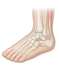
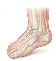
Congenital Talipes Equinovarus (Clubfoot) — Overview
Congenital Talipes Equinovarus (CTEV / Congenital Clubfoot) is a complex three-dimensional idiopathic structural deformity of the infant foot and lower leg (~1–2 per 1,000 live births, 50% bilateral, 2:1 male predominance) defined by the 4-Component 'CAVE' Deformity: Cavus (plantarflexed 1st ray creating a high medial arch), Adductus (forefoot adducted at the talonavicular/midtarsal joint), Varus (hindfoot calcaneal inversion/varus), and Equinus (severe ankle plantarflexion with shortened Achilles tendon). THE PONSETI GOLD STANDARD: The Ponseti Method (weekly gentle manipulation + above-knee long-leg serial casting for 5–6 weeks correcting components in the strict order of C -> A -> V -> E, followed by percutaneous Achilles tenotomy in 85% and Foot Abduction Brace [Denis Browne Bar] wear) achieves >95% functional, pain-free, plantigrade feet without extensive joint-scarring surgery!
Congenital Talipes Equinovarus (congenital clubfoot / PEV) is a birth condition in which one or both of a baby's feet are turned inward and downward (CAVE: high arch, forefoot turned in, heel turned in, and pointed toe). Thanks to the Ponseti Method (5–6 gentle weekly casts followed by a nighttime shoe-bar brace and daily playful stretching), over 95% of children with clubfoot run, jump, play sports, and wear normal shoes just like their peers!
Primary Causes & Biomechanics
Key Points You Should Know
- •Genetic-developmental myofibroblastic retraction in the posteromedial tarsal ligaments, tibialis posterior, flexor digitorum/hallucis longus, and Achilles tendon
- •Ponseti's Kinematic Law: The calcaneus, navicular, and cuboid are mechanically coupled and rotate together as a single unit (acetabulum pedis) around the HEAD OF THE TALUS! Therefore, the pivot point during manipulation is ALWAYS the LATERAL HEAD OF THE TALUS—NEVER touch the calcaneocuboid joint (Kite's error, which blocks hindfoot varus correction!) and NEVER pronate the forefoot (which worsens cavus!)
- •Relapse Prevention Rule: Non-adherence to the Foot Abduction Brace (Denis Browne Bar: 23h/day for 3 months, then naps + 12–14h/night until age 4–5 years) carries an 80% relapse rate, whereas strict brace adherence reduces relapse to <6%!
- •Rule #1 — The Night Brace (Denis Browne Bar) Is the Key to Keeping the Foot Straight! Casts straighten the foot in just a few weeks, but the fast-growing tendons want to pull back inward until age 4–5. Wearing the Foot Abduction Brace faithfully (23 hours/day for the first 3 months, then only during naps and bedtime until age 4–5) is the single most important secret to preventing relapse!
- •Tickle the Outer Edge of the Foot! Gently tickling or stroking from the little toe up the outer ankle encourages your baby to actively pull their foot outward and upward, strengthening the exact muscles that keep clubfoot away.
- •Flat-Heel Squatting Play for Toddlers: Once your child is walking, placing toys on the floor so they squat down with their heels flat on the ground stretches their Achilles tendons naturally all day long.
Assessment Methods
1. Goniometric ROM & Postural / Kinematic Analysis
Pirani Clubfoot Severity Score (0 to 6 points: 3 Hindfoot Contracture signs [Posterior Crease, Empty Heel, Rigid Equinus] + 3 Midfoot Contracture signs [Medial Crease, Curved Lateral Border, Lateral Head of Talus coverage]) or Dimeglio Score (0–20)**.
2. Special Clinical Provocation & Neurological/Functional Tests
Ankle dorsiflexion (target ≥15–20° post-tenotomy), forefoot abduction (target 60–70° external rotation in brace), and active peroneal eversion reflex (tickling the outer border of the foot).
3. Standardized Clinical Outcome Scales & Imaging Criteria
Pirani Score (0–6), Dimeglio Score, and ankle dorsiflexion goniometry.
1. Range of Motion & Posture Check
Your physiotherapist gently evaluates how comfortably and smoothly your pediatric subtalar, talonavicular, calcaneocuboid, and talocrural joints, plus Achilles/tibialis posterior tendons (Congenital Talipes Equinovarus — Clubfoot) moves and checks your overall posture.
2. Muscle Strength & Functional Movement Check
Your muscle balance, walking or reaching comfort, and specific gentle movement tests are evaluated without triggering sharp pain.
3. Typical Symptoms & Daily Impact Review
Foot turned downward and inward at birth (the sole of the foot faces medially/upward like a golf club head) and cannot be passively straightened to neutral on Day 1 (distinguishing true structural clubfoot from mild positional clubfoot!).
Treatment Options & Phase-Based Protocol
✨ Enriched GIF Exercise & Precaution GuidePhase 1: Acute Symptom Modulation, Tissue Protection & Early Activation
- •Phase 1 Clinical Focus & Tissue Protection: Phase 1 — Ponseti Serial Casting & French Functional Mobilization Phase (Weeks 1–8): Weekly Ponseti manipulation (1st cast: supinate the forefoot to elevate the 1st ray and correct Cavus; casts 2–5: abduct the foot around the lateral talar head to simultaneously correct Adductus and Hindfoot Varus; final step: Equinus correction + Achilles tenotomy), plus gentle towel/manual gastro-soleus & posteromedial stretching during cast changes.
- •Pain, Edema & Neuromuscular Modulation: Targeted physical modalities, relative load management, and early protected mobility of the pediatric subtalar, talonavicular, calcaneocuboid, and talocrural joints, plus Achilles/tibialis posterior tendons (Congenital Talipes Equinovarus — Clubfoot) without provoking mechanical or inflammatory irritation.
- •Proximal & Distal Kinetic Chain Maintenance: Maintain active mobility and circulation in adjacent joints while protecting the primary healing structures in Congenital Talipes Equinovarus (PEV).
Phase 2: Progressive Mobility, Motor Control & Targeted Strengthening
- •Phase 2 Progressive Mobilization & Motor Control: Phase 2 — Foot Abduction Brace (Denis Browne Bar) & Peroneal Activation Phase (Months 3–48): Strict Foot Abduction Brace adherence (affected foot set at 60–70° external rotation and 10–15° dorsiflexion), daily passive/active ankle dorsiflexion & subtalar mobilization during the 1-hour bath/play break, and tickling/stroking the lateral border of the foot to stimulate active M. peroneus longus/brevis eversion & tibialis anterior dorsiflexion!
- •Biomechanical Alignment & Co-Contraction: Restore balanced agonist-antagonist force couples, joint centration, and multi-planar neuromuscular control across the pediatric subtalar, talonavicular, calcaneocuboid, and talocrural joints, plus Achilles/tibialis posterior tendons (Congenital Talipes Equinovarus — Clubfoot).
- •Myofascial Extensibility & Endurance: Address secondary compensatory tightness and build local muscular endurance within safe, symptom-free biomechanical parameters.
Phase 3: Functional Integration, Proprioception & Long-Term Prevention
- •Phase 3 Functional Integration & Long-Term Prevention: Phase 3 — Squatting, Plantigrade Gait & Dynamic Balance Phase (Ages 1–5+): Deep flat-heel toddler squatting play (natural daily Achilles stretch!), walking up inclined ramps, heel-walking & single-leg balance games, and monitoring for dynamic supination (tibialis anterior over-pull).
- •Proprioceptive Balance & Dynamic Loading: Progress to task-specific closed-chain, balance, and functional movement patterns tailored to daily life, occupational, or athletic demands.
- •Self-Management & Relapse Prophylaxis: Establish lifelong ergonomic habits, home exercise maintenance, and early warning-sign awareness to prevent recurrence.
Step 1: Calming Pain, Protecting the Area & Gentle Starter Motion
- •Rule #1 — The Night Brace (Denis Browne Bar) Is the Key to Keeping the Foot Straight! Casts straighten the foot in just a few weeks, but the fast-growing tendons want to pull back inward until age 4–5. Wearing the Foot Abduction Brace faithfully (23 hours/day for the first 3 months, then only during naps and bedtime until age 4–5) is the single most important secret to preventing relapse!
- •Calm Protection & Gentle Start: During the initial stage of Congenital Clubfoot, avoid painful overloading and perform the gentle, pain-free starter movements shown below to support circulation and comfort.
- •Respecting Your Body's Signals: Every movement should stay within a comfortable, non-painful zone—never force through sharp or radiating pain.
Step 2: Restoring Flexibility & Building Supporting Muscle Strength
- •Tickle the Outer Edge of the Foot! Gently tickling or stroking from the little toe up the outer ankle encourages your baby to actively pull their foot outward and upward, strengthening the exact muscles that keep clubfoot away.
- •Step-by-Step Flexibility & Support: As acute sensitivity settles, gradually build mobility and strengthen the supporting muscles around the joint at a steady, controlled pace.
- •Posture & Breathing Coordination: Keep your posture upright and breathe smoothly (never hold your breath!) during every repetition.
Step 3: Safe Daily Habits, Stamina & Preventing Recurrence
- •Flat-Heel Squatting Play for Toddlers: Once your child is walking, placing toys on the floor so they squat down with their heels flat on the ground stretches their Achilles tendons naturally all day long.
- •Confident Return to Daily Life: Integrate your stronger muscles and improved balance into walking, stairs, work, and recreational activities.
- •Preventing Future Flare-Ups: Continue your core protective habits and key maintenance exercises even after symptoms resolve.
Animated (GIF) Therapeutic Exercise Guide
Animated clinical exercise protocol with biomechanical angles, target muscle groups, and dosage parameters (Click image to enlarge)
Step-by-step animated exercise demonstrations you can safely perform at home (Click image to enlarge)
 🔍 Enlarge
1. Long-Sitting Towel-Assisted Gastrocnemius-Soleus & Plantar Fascia Stretch
1. Seated Towel Calf & Foot Arch Stretch
Long-sitting with a towel looped around the metatarsal heads, applying a slow, sustained passive pull into ankle dorsiflexion. Restores gastrocnemius-soleus-Achilles length and reduces plantar fascial and equine tension. Specifically indicated in Congenital Talipes Equinovarus (PEV) rehabilitation to optimize pediatric subtalar, talonavicular, calcaneocuboid, and talocrural joints, plus Achilles/tibialis posterior tendons (Congenital Talipes Equinovarus — Clubfoot) biomechanics and neuromuscular control.
Sit with your leg extended and loop a towel around the ball of your foot. Gently pull both ends of the towel toward you until you feel a comfortable stretch in your calf and sole, holding for 20–30 seconds. Perform smoothly within a comfortable, pain-free range to support your Congenital Clubfoot recovery.
 🔍 Enlarge
2. Rhythmic Active Ankle Dorsiflexion & Plantarflexion Pumping (Venous/Edema Pump)
2. Ankle Toe-Pulls and Pointing (Circulation & Swelling Pump)
Rhythmic alternating contraction of the tibialis anterior and gastrocnemius-soleus complex. Activates the deep venous calf pump to prevent deep vein thrombosis (DVT), clears distal edema, and maintains talocrural mobility. Specifically indicated in Congenital Talipes Equinovarus (PEV) rehabilitation to optimize pediatric subtalar, talonavicular, calcaneocuboid, and talocrural joints, plus Achilles/tibialis posterior tendons (Congenital Talipes Equinovarus — Clubfoot) biomechanics and neuromuscular control.
While lying down or sitting with your legs supported, pull your toes and feet firmly upward toward your shins, hold for 3 seconds, then point them downward like pressing a gas pedal. Perform smoothly within a comfortable, pain-free range to support your Congenital Clubfoot recovery.
 🔍 Enlarge
3. Intrinsic Foot Core ('Short Foot') & Medial Longitudinal Arch Activation
3. Seated Arch Doming (Short Foot) & Towel Scrunch
Drawing the first metatarsal head toward the calcaneus to elevate the medial longitudinal arch without curling the toes (or performing controlled towel curls). Strengthens the intrinsic plantar muscles supporting the subtalar and midtarsal joints. Specifically indicated in Congenital Talipes Equinovarus (PEV) rehabilitation to optimize pediatric subtalar, talonavicular, calcaneocuboid, and talocrural joints, plus Achilles/tibialis posterior tendons (Congenital Talipes Equinovarus — Clubfoot) biomechanics and neuromuscular control.
While seated with your foot flat on the floor (or on a small towel), gently draw the ball of your foot toward your heel to lift your inner arch without curling your toes, hold for 5 seconds, and relax. Perform smoothly within a comfortable, pain-free range to support your Congenital Clubfoot recovery.
 🔍 Enlarge
4. Functional Closed-Kinetic-Chain Chair Transfer (Sit-to-Stand Biomechanics)
4. Controlled Chair Sit-to-Stand Transfer
Controlled transition from sitting to standing and eccentric lowering back to the chair with symmetrical weight distribution, anterior trunk inclination ('nose over toes'), and neutral frontal knee alignment. Specifically indicated in Congenital Talipes Equinovarus (PEV) rehabilitation to optimize pediatric subtalar, talonavicular, calcaneocuboid, and talocrural joints, plus Achilles/tibialis posterior tendons (Congenital Talipes Equinovarus — Clubfoot) biomechanics and neuromuscular control.
Sit near the front of a sturdy chair with your feet hip-width apart. Lean slightly forward and push evenly through both heels to stand up tall, then slowly lower yourself back down over 3 seconds. Perform smoothly within a comfortable, pain-free range to support your Congenital Clubfoot recovery.
 🔍 Enlarge
5. Supported Standing Hip Abduction & Closed-Chain Stance Stabilization
5. Counter-Supported Standing Side Leg Lift
Standing upright with light hand support, abducting the lower limb in the frontal plane while the stance-leg gluteus medius contracts isometrically to keep the pelvis level. Builds functional gait stability. Specifically indicated in Congenital Talipes Equinovarus (PEV) rehabilitation to optimize pediatric subtalar, talonavicular, calcaneocuboid, and talocrural joints, plus Achilles/tibialis posterior tendons (Congenital Talipes Equinovarus — Clubfoot) biomechanics and neuromuscular control.
Hold lightly onto a sturdy counter or chair back and stand tall. Without leaning your trunk sideways, lift your leg out to the side, pause for 2–3 seconds, and slowly return. Perform smoothly within a comfortable, pain-free range to support your Congenital Clubfoot recovery.
 🔍 Enlarge
6. Closed-Chain Bilateral/Eccentric Heel Raise (Triceps Surae & Achilles Loading)
6. Supported Standing Heel Raise & Slow Lowering
Controlled concentric heel elevation followed by slow (3–4 second) eccentric lowering into neutral dorsiflexion. Stimulates Type I collagen alignment in the Achilles/plantar aponeurosis and builds ankle plantarflexor propulsion. Specifically indicated in Congenital Talipes Equinovarus (PEV) rehabilitation to optimize pediatric subtalar, talonavicular, calcaneocuboid, and talocrural joints, plus Achilles/tibialis posterior tendons (Congenital Talipes Equinovarus — Clubfoot) biomechanics and neuromuscular control.
Hold lightly onto a wall or chair back. Rise up onto the balls of both feet, pause at the top, and slowly lower your heels back down to the floor over 3–4 seconds. Perform smoothly within a comfortable, pain-free range to support your Congenital Clubfoot recovery.
Movements to Avoid & Strict Precautions
1. Stopping the Nighttime Foot Abduction Brace Early Because the Foot 'Looks Completely Normal' (80% Relapse Rate!)
1. Stopping the bedtime shoe-bar brace before age 4–5 just because the foot looks straight (this is the #1 cause of clubfoot coming back!)
2. Leaving a Slipped Cast On (Causes Severe Pressure Sores on the Top of the Foot!)
2. Leaving a cast on if the baby's toes have slid inward out of sight
3. Pushing Up Under the Metatarsal Heads Before Forefoot Abduction Is Corrected (Creates a Rocker-Bottom Foot!)
3. Forcefully bending the front of the foot upward before the inward turn is corrected
4. Buying Special Stiff Orthopedic Boots Instead of Normal Flexible Sneakers for Daytime Walking
4. Once in the night-brace phase, children wear completely normal, comfortable daytime sneakers—not heavy orthopedic boots!
Congenital Talipes Equinovarus (Clubfoot) — Video Guide
Click directly on any video to open and watch in a window, or review details via 📋 Summary & Options.
Condition Overview Video (1 Video)
Summary of pathophysiology, biomechanics, and clinical presentation
What the condition is, why it develops, and its basic mechanism

What Is Congenital Clubfoot (Talipes Equinovarus)? Ponseti Method Explained
Congenital Talipes Equinovarus (CTEV / Congenital Clubfoot) is a complex three-dimensional idiopathic structural deformity of the infant foot and lower leg (~1–2 per 1,000 live births, 50% bilateral, 2:1 male predominance) defined by the 4-Component 'CAVE' Deformity: Cavus (plantarflexed 1st ray creating a high medial arch), Adductus (forefoot adducted at the talonavicular/midtarsal joint), Varus (hindfoot calcaneal inversion/varus), and Equinus (severe ankle plantarflexion with shortened Achilles tendon). THE PONSETI GOLD STANDARD: The Ponseti Method (weekly gentle manipulation + above-knee long-leg serial casting for 5–6 weeks correcting components in the strict order of C -> A -> V -> E, followed by percutaneous Achilles tenotomy in 85% and Foot Abduction Brace [Denis Browne Bar] wear) achieves >95% functional, pain-free, plantigrade feet without extensive joint-scarring surgery!
Congenital Talipes Equinovarus (congenital clubfoot / PEV) is a birth condition in which one or both of a baby's feet are turned inward and downward (CAVE: high arch, forefoot turned in, heel turned in, and pointed toe). Thanks to the Ponseti Method (5–6 gentle weekly casts followed by a nighttime shoe-bar brace and daily playful stretching), over 95% of children with clubfoot run, jump, play sports, and wear normal shoes just like their peers!
Treatment and Exercise Videos
Phase-based physiotherapy protocols, mobilization, and therapeutic exercises
Step-by-step recovery and safe home exercise applications
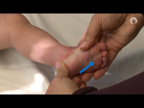
Clubfoot (Talipes Equinovarus) Stretching, Mobilization, and Massage Techniques
Phase 1: Acute Symptom Modulation, Tissue Protection & Early Activation: Phase 1 Clinical Focus & Tissue Protection: Phase 1 — Ponseti Serial Casting & French Functional Mobilization Phase (Weeks 1–8): Weekly Ponseti manipulation (1st cast: supinate the forefoot to elevate the 1st ray and correct Cavus; casts 2–5: abduct the foot around the lateral talar head to simultaneously correct Adductus and Hindfoot Varus; final step: Equinus correction + Achilles tenotomy), plus gentle towel/manual gastro-soleus & posteromedial stretching during cast changes.
Step 1: Calming Pain, Protecting the Area & Gentle Starter Motion: Rule #1 — The Night Brace (Denis Browne Bar) Is the Key to Keeping the Foot Straight! Casts straighten the foot in just a few weeks, but the fast-growing tendons want to pull back inward until age 4–5. Wearing the Foot Abduction Brace faithfully (23 hours/day for the first 3 months, then only during naps and bedtime until age 4–5) is the single most important secret to preventing relapse!

Mobilization and Physiotherapy Techniques in Pediatric Foot Deformities
Phase 2: Progressive Mobility, Motor Control & Targeted Strengthening: Phase 2 Progressive Mobilization & Motor Control: Phase 2 — Foot Abduction Brace (Denis Browne Bar) & Peroneal Activation Phase (Months 3–48): Strict Foot Abduction Brace adherence (affected foot set at 60–70° external rotation and 10–15° dorsiflexion), daily passive/active ankle dorsiflexion & subtalar mobilization during the 1-hour bath/play break, and tickling/stroking the lateral border of the foot to stimulate active M. peroneus longus/brevis eversion & tibialis anterior dorsiflexion!
Step 2: Restoring Flexibility & Building Supporting Muscle Strength: Tickle the Outer Edge of the Foot! Gently tickling or stroking from the little toe up the outer ankle encourages your baby to actively pull their foot outward and upward, strengthening the exact muscles that keep clubfoot away.
Clinical Assessment, Goniometry & Special Test Videos
Displayed only in Healthcare Professional mode (hidden in Patient mode)

Ankle and Foot Goniometric Measurement (Dorsiflexion, Plantarflexion, Inversion/Eversion)
Goniometric ROM & Postural / Kinematic Analysis: Pirani Clubfoot Severity Score (0 to 6 points: 3 Hindfoot Contracture signs [Posterior Crease, Empty Heel, Rigid Equinus] + 3 Midfoot Contracture signs [Medial Crease, Curved Lateral Border, Lateral Head of Talus coverage]) or Dimeglio Score (0–20).
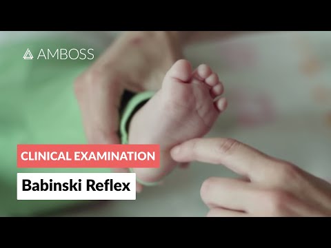
Pediatric Neurological Examination: Babinski and Plantar Reflex Assessment
Special Clinical Provocation & Neurological/Functional Tests: Ankle dorsiflexion (target ≥15–20° post-tenotomy), forefoot abduction (target 60–70° external rotation in brace), and active peroneal eversion reflex (tickling the outer border of the foot).