Congenital Muscular TorticollisCongenital Wry Neck (Congenital Muscular Torticollis)
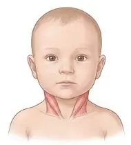
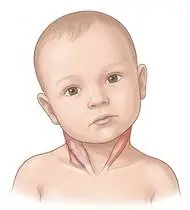
Congenital Muscular Torticollis — Overview
Congenital Muscular Torticollis (CMT — 'Infant Wry Neck') is the third most common congenital orthopedic anomaly in infants, caused by unilateral shortening, fibrosis, or a benign fibrotic pseudotumor (fibromatosis colli, palpable at weeks 2–6 in the lower third of the muscle) of the M. sternocleidomastoid (SCM). Because the SCM originates on the sternum/clavicle and inserts on the ipsilateral mastoid process, contraction/shortening of one SCM pulls the head into IPSILATERAL LATERAL FLEXION (ear tilted toward the tight muscle) and CONTRALATERAL ROTATION (chin/face turned toward the OPPOSITE shoulder)! Clinical evidence proves that when physiotherapy starts BEFORE 1 to 3 months of age, >95–98% of infants achieve 100% complete resolution without surgery while preventing deformational plagiocephaly (flat head) and facial asymmetry!
Congenital muscular torticollis (infant wry neck) occurs when the neck muscle on one side (sternocleidomastoid / SCM) is tight or shortened at birth—causing your baby to tilt their ear toward the tight side while turning their chin to look toward the opposite side. The wonderful news is that when started in the first months of life, gentle diaper-change stretches, smart carrying positions, and tummy time cure over 95% of babies completely without surgery!
Primary Causes & Biomechanics
Key Points You Should Know
- •Intrauterine crowding (primigravida, breech, twins, oligohydramnios) causing compartment ischemia or birth microtrauma of the unilateral SCM
- •Biomechanics of the SCM (Opposite Tilt & Turn!): Right CMT = Head tilted to the Right + Chin rotated to the Left; Left CMT = Head tilted to the Left + Chin rotated to the Right
- •Secondary Consequences if Untreated: Positional Plagiocephaly (flattening of the contralateral occiput from always sleeping looking to one side), craniofacial hemihypoplasia, and C-shaped developmental cervical/thoracic scoliosis
- •Understanding the Tilt vs. Turn Rule: If the RIGHT neck muscle is tight, your baby tilts their right ear down toward the right shoulder, but turns their chin to look to the LEFT! That means to help them, we place all toys, your face, and the bottle on the RIGHT side so they turn their chin to look right, and we gently tilt their left ear toward the left shoulder.
- •The Magic 'Side-Hold' Carrying Trick: Hold your baby on their side across your arms with the TIGHT neck side facing DOWN against your forearm. This single position stretches the tight bottom muscle with gravity *and* strengthens the weak top muscle whenever your baby looks around!
- •60 Minutes of Daily Tummy Time (Split Up): Tummy time takes pressure off the flat spot on the back of the head (plagiocephaly) and builds balanced neck strength.
Assessment Methods
1. Goniometric ROM & Postural / Kinematic Analysis
Cervical passive and active Rotation (N: 100–110° in infants — chin past the acromion) and Lateral Flexion (N: 65–70° — ear to shoulder) measured via arthrodial protractor/goniometry.
2. Special Clinical Provocation & Neurological/Functional Tests
Muscle Function Scale (MFS 0–5) (holding the infant horizontally in lateral suspension and scoring active antigravity head righting of the *healthy* contralateral SCM!), palpation of the SCM for a fibromatosis colli olive-sized nodule, Craniofacial Plagiocephaly Severity Scale, and mandatory Hip Screening (5–10% of CMT infants have concomitant Hip Dysplasia [DDH]!).
3. Standardized Clinical Outcome Scales & Imaging Criteria
Cervical Rotation & Lateral Flexion Deficit (degrees, Kaplan Severity Grades 1–8), Muscle Function Scale (MFS), and Cranial Vault Asymmetry Index.
1. Range of Motion & Posture Check
Your physiotherapist gently evaluates how comfortably and smoothly your unilateral Sternocleidomastoid (SCM) muscle (fibromatosis colli / shortening) and craniocervical alignment moves and checks your overall posture.
2. Muscle Strength & Functional Movement Check
Your muscle balance, walking or reaching comfort, and specific gentle movement tests are evaluated without triggering sharp pain.
3. Typical Symptoms & Daily Impact Review
Consistent Head Tilt & Chin Preference: The baby consistently tilts their head to one side (ear toward shoulder) and prefers looking exclusively toward the opposite side (especially noticeable in photos!).
Treatment Options & Phase-Based Protocol
✨ Enriched GIF Exercise & Precaution GuidePhase 1: Acute Symptom Modulation, Tissue Protection & Early Activation
- •Phase 1 Clinical Focus & Tissue Protection: Phase 1 — Two-Person / Scapula-Stabilized Gentle SCM Stretch & Positioning Phase: Stretch towards the RESTRICTED directions (Rotate the chin TOWARD the tight side; Tilt the ear AWAY from the tight side toward the opposite shoulder!) gently and painlessly at every diaper change (while stabilizing the ipsilateral shoulder down!), and arrange the crib, feeding bottle/breast, and colorful toys on the TIGHT side so the baby actively turns their chin toward the tight side all day!
- •Pain, Edema & Neuromuscular Modulation: Targeted physical modalities, relative load management, and early protected mobility of the unilateral Sternocleidomastoid (SCM) muscle (fibromatosis colli / shortening) and craniocervical alignment without provoking mechanical or inflammatory irritation.
- •Proximal & Distal Kinetic Chain Maintenance: Maintain active mobility and circulation in adjacent joints while protecting the primary healing structures in Congenital Muscular Torticollis.
Phase 2: Progressive Mobility, Motor Control & Targeted Strengthening
- •Phase 2 Progressive Mobilization & Motor Control: Phase 2 — Side-Lying 'Football Carry' & Contralateral SCM Strengthening Phase: Carry the infant in the 'Football / Side-Lying Carry' with the TIGHT SCM SIDE DOWN against your forearm (gravity gently stretches the bottom tight SCM while the baby actively lifts their head upward using the weak upper SCM!), plus 30–60 min/day of supervised Tummy Time.
- •Biomechanical Alignment & Co-Contraction: Restore balanced agonist-antagonist force couples, joint centration, and multi-planar neuromuscular control across the unilateral Sternocleidomastoid (SCM) muscle (fibromatosis colli / shortening) and craniocervical alignment.
- •Myofascial Extensibility & Endurance: Address secondary compensatory tightness and build local muscular endurance within safe, symptom-free biomechanical parameters.
Phase 3: Functional Integration, Proprioception & Long-Term Prevention
- •Phase 3 Functional Integration & Long-Term Prevention: Phase 3 — Active Optical Righting, Rolling & Midline Symmetry Phase: Visual tracking games rotating the head past midline toward the affected side, symmetrical rolling to both sides, and sitting midline head control.
- •Proprioceptive Balance & Dynamic Loading: Progress to task-specific closed-chain, balance, and functional movement patterns tailored to daily life, occupational, or athletic demands.
- •Self-Management & Relapse Prophylaxis: Establish lifelong ergonomic habits, home exercise maintenance, and early warning-sign awareness to prevent recurrence.
Step 1: Calming Pain, Protecting the Area & Gentle Starter Motion
- •Understanding the Tilt vs. Turn Rule: If the RIGHT neck muscle is tight, your baby tilts their right ear down toward the right shoulder, but turns their chin to look to the LEFT! That means to help them, we place all toys, your face, and the bottle on the RIGHT side so they turn their chin to look right, and we gently tilt their left ear toward the left shoulder.
- •Calm Protection & Gentle Start: During the initial stage of Congenital Wry Neck, avoid painful overloading and perform the gentle, pain-free starter movements shown below to support circulation and comfort.
- •Respecting Your Body's Signals: Every movement should stay within a comfortable, non-painful zone—never force through sharp or radiating pain.
Step 2: Restoring Flexibility & Building Supporting Muscle Strength
- •The Magic 'Side-Hold' Carrying Trick: Hold your baby on their side across your arms with the TIGHT neck side facing DOWN against your forearm. This single position stretches the tight bottom muscle with gravity *and* strengthens the weak top muscle whenever your baby looks around!
- •Step-by-Step Flexibility & Support: As acute sensitivity settles, gradually build mobility and strengthen the supporting muscles around the joint at a steady, controlled pace.
- •Posture & Breathing Coordination: Keep your posture upright and breathe smoothly (never hold your breath!) during every repetition.
Step 3: Safe Daily Habits, Stamina & Preventing Recurrence
- •60 Minutes of Daily Tummy Time (Split Up): Tummy time takes pressure off the flat spot on the back of the head (plagiocephaly) and builds balanced neck strength.
- •Confident Return to Daily Life: Integrate your stronger muscles and improved balance into walking, stairs, work, and recreational activities.
- •Preventing Future Flare-Ups: Continue your core protective habits and key maintenance exercises even after symptoms resolve.
Animated (GIF) Therapeutic Exercise Guide
Animated clinical exercise protocol with biomechanical angles, target muscle groups, and dosage parameters (Click image to enlarge)
Step-by-step animated exercise demonstrations you can safely perform at home (Click image to enlarge)
 🔍 Enlarge
1. Submaximal Cervical Lateral Flexion & Upper Trapezius / Scalene Myofascial Stretch
1. Gentle Side Neck & Shoulder Relaxation Stretch
With the ipsilateral shoulder depressed and stabilized, gently tilting the head toward the opposite shoulder within a strictly pain-free, non-radicular range. Releases hypertonic upper trapezius, levator scapulae, and scalene myofascial trigger points. Specifically indicated in Congenital Muscular Torticollis rehabilitation to optimize unilateral Sternocleidomastoid (SCM) muscle (fibromatosis colli / shortening) and craniocervical alignment biomechanics and neuromuscular control.
Sit upright and relax your shoulders down. Slowly tilt your ear toward your opposite shoulder until you feel a gentle, comfortable stretch along the side of your neck, hold for 20 seconds while breathing calmly, and return to center. Perform smoothly within a comfortable, pain-free range to support your Congenital Wry Neck recovery.
 🔍 Enlarge
2. Prone Positioning & Controlled Thoracolumbar Extension (McKenzie / Back Extensor)
2. Prone Lying & Gentle Back Extension Lift
Prone positioning and graduated thoracic/lumbar extension (with a pillow under the abdomen when indicated for spinal unloading). Counters flexed postural kyphosis, stretches tight hip flexors, and strengthens paraspinal extensors. Specifically indicated in Congenital Muscular Torticollis rehabilitation to optimize unilateral Sternocleidomastoid (SCM) muscle (fibromatosis colli / shortening) and craniocervical alignment biomechanics and neuromuscular control.
Lie on your stomach (place a small pillow under your hips/tummy if needed for comfort). Gently lift your head and chest slightly off the mat by squeezing your back muscles without straining your neck, hold for 3–5 seconds, and lower. Perform smoothly within a comfortable, pain-free range to support your Congenital Wry Neck recovery.
 🔍 Enlarge
3. Scapulothoracic Retraction & Depression (Middle/Lower Trapezius & Rhomboids)
3. Shoulder Blade Squeeze (Postural Alignment)
Adduction and depression of the scapulae via the middle/lower trapezius and rhomboids while inhibiting upper trapezius overactivity. Corrects anterior scapular tilt, widens the subacromial outlet, and unloads the anterior thoracic column. Specifically indicated in Congenital Muscular Torticollis rehabilitation to optimize unilateral Sternocleidomastoid (SCM) muscle (fibromatosis colli / shortening) and craniocervical alignment biomechanics and neuromuscular control.
Sit or stand tall. Keep your shoulders relaxed away from your ears, gently draw your shoulder blades together and slightly downward toward your back pockets, hold for 5 seconds, and release. Perform smoothly within a comfortable, pain-free range to support your Congenital Wry Neck recovery.
 🔍 Enlarge
4. Craniocervical Axial Retraction ('Chin-Tuck' — Deep Cervical Flexor Activation)
4. Chin-Tuck (Double-Chin) Postural Neck Alignment
Horizontal posterior translation of the cranium and cervical spine without tipping the head up or down. Activates the deep cervical flexors (longus colli/capitis), decompresses suboccipital structures, and opens the lower cervical intervertebral foramina. Specifically indicated in Congenital Muscular Torticollis rehabilitation to optimize unilateral Sternocleidomastoid (SCM) muscle (fibromatosis colli / shortening) and craniocervical alignment biomechanics and neuromuscular control.
Sit or stand tall looking straight ahead. Gently glide your chin straight backward (as if making a subtle double chin) without tilting your head down, feel the back of your neck lengthen, hold for 5 seconds, and relax. Perform smoothly within a comfortable, pain-free range to support your Congenital Wry Neck recovery.
 🔍 Enlarge
5. Bobath Bilateral Clasp-Hand Elevation & Midline Trunk Organization
5. Clasped-Hands Two-Arm Overhead Reach
Interlocking the fingers with the affected thumb positioned on top, maintaining elbow extension and scapular protraction while elevating both arms symmetrically in the midline. Inhibits upper limb flexor synergy and prevents hemiplegic shoulder impingement. Specifically indicated in Congenital Muscular Torticollis rehabilitation to optimize unilateral Sternocleidomastoid (SCM) muscle (fibromatosis colli / shortening) and craniocervical alignment biomechanics and neuromuscular control.
Clasp your hands together with your affected thumb on top. Keeping both elbows straight, use your stronger arm to smoothly guide both hands upward toward the ceiling and slowly back down. Perform smoothly within a comfortable, pain-free range to support your Congenital Wry Neck recovery.
 🔍 Enlarge
6. Quadruped Contralateral Arm-Leg Reach ('Bird-Dog' Multifidus & Deep Core)
6. All-Fours Opposite Arm & Leg Reach (Bird-Dog)
In quadruped with a neutral lumbopelvic spine and abdominal drawing-in (transversus abdominis), extending opposite upper and lower extremities horizontally without pelvic rotation or lumbar hyperextension. Minimizes spinal compressive load while maximizing multifidus recruitment. Specifically indicated in Congenital Muscular Torticollis rehabilitation to optimize unilateral Sternocleidomastoid (SCM) muscle (fibromatosis colli / shortening) and craniocervical alignment biomechanics and neuromuscular control.
On your hands and knees, keep your back flat like a tabletop and gently tighten your tummy. Slowly reach one arm forward and the opposite leg straight back withoutarching your lower back, hold for 5 seconds, and switch sides. Perform smoothly within a comfortable, pain-free range to support your Congenital Wry Neck recovery.
Movements to Avoid & Strict Precautions
1. Forcing Neck Stretches While the Baby Is Crying and Stiffening (Causes Muscle Micro-Tears & Fibrosis!)
1. Forcing the baby's neck while they are crying and fighting the stretch (always calm the baby first and stretch gently!)
2. Leaving the Baby in Car Seats, Bouncers, or Swings for Hours (Worsens Flat Head & Head Tilt!)
2. Leaving the baby lying in car seats, swings, or strollers at home (use flat floor tummy-time instead!)
3. Feeding and Approaching the Baby From Their 'Easy/Favorite' Side (Reinforces the Tightness!)
3. Standing and feeding from the side the baby already likes to look toward (always approach from the tight side!)
4. Rotating the Baby's Head Without Gently Holding Down the Shoulder on the Tight Side
4. Turning the baby's head while letting their shoulder hike up to follow the chin
Congenital Muscular Torticollis — Video Guide
Click directly on any video to open and watch in a window, or review details via 📋 Summary & Options.
Condition Overview Video (1 Video)
Summary of pathophysiology, biomechanics, and clinical presentation
What the condition is, why it develops, and its basic mechanism

What Is Congenital Muscular Torticollis (Infant Wry Neck)? How to Recognize It
Congenital Muscular Torticollis (CMT — 'Infant Wry Neck') is the third most common congenital orthopedic anomaly in infants, caused by unilateral shortening, fibrosis, or a benign fibrotic pseudotumor (fibromatosis colli, palpable at weeks 2–6 in the lower third of the muscle) of the M. sternocleidomastoid (SCM). Because the SCM originates on the sternum/clavicle and inserts on the ipsilateral mastoid process, contraction/shortening of one SCM pulls the head into IPSILATERAL LATERAL FLEXION (ear tilted toward the tight muscle) and CONTRALATERAL ROTATION (chin/face turned toward the OPPOSITE shoulder)! Clinical evidence proves that when physiotherapy starts BEFORE 1 to 3 months of age, >95–98% of infants achieve 100% complete resolution without surgery while preventing deformational plagiocephaly (flat head) and facial asymmetry!
Congenital muscular torticollis (infant wry neck) occurs when the neck muscle on one side (sternocleidomastoid / SCM) is tight or shortened at birth—causing your baby to tilt their ear toward the tight side while turning their chin to look toward the opposite side. The wonderful news is that when started in the first months of life, gentle diaper-change stretches, smart carrying positions, and tummy time cure over 95% of babies completely without surgery!
Treatment and Exercise Videos
Phase-based physiotherapy protocols, mobilization, and therapeutic exercises
Step-by-step recovery and safe home exercise applications

Infant Torticollis Exercises | Safe Stretching and Positioning for Wry Neck
Phase 1: Acute Symptom Modulation, Tissue Protection & Early Activation: Phase 1 Clinical Focus & Tissue Protection: Phase 1 — Two-Person / Scapula-Stabilized Gentle SCM Stretch & Positioning Phase: Stretch towards the RESTRICTED directions (Rotate the chin TOWARD the tight side; Tilt the ear AWAY from the tight side toward the opposite shoulder!) gently and painlessly at every diaper change (while stabilizing the ipsilateral shoulder down!), and arrange the crib, feeding bottle/breast, and colorful toys on the TIGHT side so the baby actively turns their chin toward the tight side all day!
Step 1: Calming Pain, Protecting the Area & Gentle Starter Motion: Understanding the Tilt vs. Turn Rule: If the RIGHT neck muscle is tight, your baby tilts their right ear down toward the right shoulder, but turns their chin to look to the LEFT! That means to help them, we place all toys, your face, and the bottle on the RIGHT side so they turn their chin to look right, and we gently tilt their left ear toward the left shoulder.

Torticollis Exercises, Carrying Positions, and Tummy Time Play Tips
Phase 2: Progressive Mobility, Motor Control & Targeted Strengthening: Phase 2 Progressive Mobilization & Motor Control: Phase 2 — Side-Lying 'Football Carry' & Contralateral SCM Strengthening Phase: Carry the infant in the 'Football / Side-Lying Carry' with the TIGHT SCM SIDE DOWN against your forearm (gravity gently stretches the bottom tight SCM while the baby actively lifts their head upward using the weak upper SCM!), plus 30–60 min/day of supervised Tummy Time.
Step 2: Restoring Flexibility & Building Supporting Muscle Strength: The Magic 'Side-Hold' Carrying Trick: Hold your baby on their side across your arms with the TIGHT neck side facing DOWN against your forearm. This single position stretches the tight bottom muscle with gravity *and* strengthens the weak top muscle whenever your baby looks around!
Clinical Assessment, Goniometry & Special Test Videos
Displayed only in Healthcare Professional mode (hidden in Patient mode)
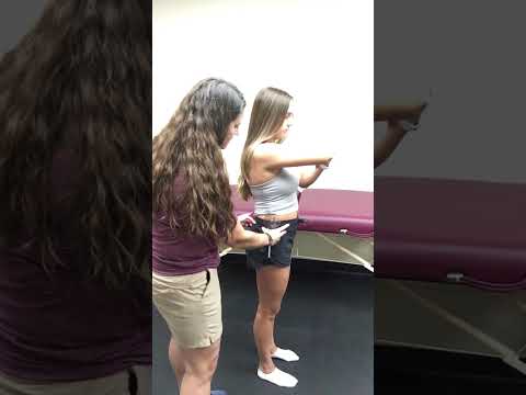
Vertebral Column (Cervical & Thoracolumbar Spine) Goniometric and Tape Measurement
Goniometric ROM & Postural / Kinematic Analysis: Cervical passive and active Rotation (N: 100–110° in infants — chin past the acromion) and Lateral Flexion (N: 65–70° — ear to shoulder) measured via arthrodial protractor/goniometry.

Newborn and Infant Primitive Reflex Examination (Moro, Grasp, ATNR, Galant)
Special Clinical Provocation & Neurological/Functional Tests: Muscle Function Scale (MFS 0–5) (holding the infant horizontally in lateral suspension and scoring active antigravity head righting of the *healthy* contralateral SCM!), palpation of the SCM for a fibromatosis colli olive-sized nodule, Craniofacial Plagiocephaly Severity Scale, and mandatory Hip Screening (5–10% of CMT infants have concomitant Hip Dysplasia [DDH]!).