Charcot-Marie-Tooth (CMT) DiseaseHereditary Hand & Foot Nerve Wasting (Charcot-Marie-Tooth Disease (CMT))
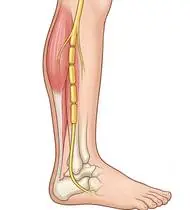
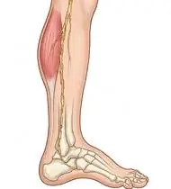
Charcot-Marie-Tooth (CMT) Disease — Overview
Charcot-Marie-Tooth Disease (CMT / Hereditary Motor and Sensory Neuropathy [HMSN]) is the most common inherited neurological disorder (~1 in 2,500 people), caused by mutations in peripheral nerve myelin or axonal proteins (CMT1A: PMP22 duplication on chromosome 17p causing slow demyelinating NCV <38 m/s [~60–70% of cases]; CMT2: mitofusin-2 axonal degeneration). It produces a slowly progressive, symmetric, length-dependent DISTAL > PROXIMAL motor and sensory neuropathy starting in childhood/adolescence (ages 5–20). THE BIOMECHANICAL HALLMARK OF CMT: Selective early denervation of the intrinsic foot muscles, M. tibialis anterior (dorsiflexor), and M. peroneus brevis (evertor) while the stronger M. tibialis posterior (invertor) and M. peroneus longus (which plantarflexes the 1st metatarsal head!) remain relatively preserved drives the foot into a progressive Pes Cavovarus Deformity (high arch + forefoot pronation + hindfoot varus + claw toes) alongside 'Stork-Leg / Inverted Champagne Bottle' distal calf atrophy and Steppage Gait!
Charcot-Marie-Tooth disease (CMT / hereditary hand & foot nerve condition) is an inherited condition affecting the peripheral nerves traveling to the feet/ankles and hands, usually starting in childhood or the teen years. Because CMT progresses very slowly and never affects the brain or lifespan, combining daily calf/arch stretching, lightweight carbon ankle supports or shoe inserts, hip-strengthening balance exercises, and fine hand exercises keeps children and adults active, steady, and independent for life!
Primary Causes & Biomechanics
Key Points You Should Know
- •Length-dependent hereditary demyelination (CMT1) or axonal loss (CMT2) affecting the longest motor and sensory nerves to the feet and hands
- •The Peroneus Longus – Peroneus Brevis Force-Couple Imbalance: Strong peroneus longus plantarflexes the 1st ray while weak tibialis anterior & peroneus brevis fail to lift/evert the foot—creating a 1st-ray-driven Cavovarus deformity and recurrent lateral ankle sprains!
- •Late-onset distal intrinsic hand muscle atrophy (interossei, lumbricals, thenar) impairing pinch, buttoning, and handwriting
- •Why the Foot Arch Gets High (Pes Cavus) and Ankles Roll Outward: In CMT, some ankle muscles stay strong while others weaken, pulling the base of the big toe downward and tilting the heel inward. A simple custom shoe insole (with a recess under the big toe and outer wedge) or a feather-light carbon ankle brace (AFO) stops ankle sprains and tripping immediately!
- •Train Your Hips to Steady Your Ankles: When the ankle muscles are weak, your hip and thigh muscles become your body's main balance stabilizers—keeping your hips strong with bridges and side leg lifts stops stumbles.
- •Paced Exercise (Never to Total Exhaustion): Moderate swimming, cycling, and submaximal strength training are great for CMT, as long as you avoid exhausting overwork.
Assessment Methods
1. Goniometric ROM & Postural / Kinematic Analysis
CMTPedS (CMT Pediatric Scale, 0–44) / CMTNS (CMT Neuropathy Score), ankle dorsiflexion/plantarflexion/eversion/inversion MMT, and Foot Posture Index (Pes Cavus).
2. Special Clinical Provocation & Neurological/Functional Tests
Coleman Lateral Block Test (GOLD STANDARD test to determine if hindfoot varus in a Pes Cavovarus foot is *flexible* and driven by the plantarflexed 1st ray: placing the heel and lateral foot on a 2.5 cm block while letting the 1st metatarsal head hang free to the floor—if the heel corrects to neutral, it is flexible and responds to a 1st-ray cutout + lateral wedge orthotic!), plus 6MWT and Jamar/pinch dynamometry.
3. Standardized Clinical Outcome Scales & Imaging Criteria
CMTPedS / CMTNS, Coleman Block Test flexibility, 6MWT, and NCV/genetic testing.
1. Range of Motion & Posture Check
Your physiotherapist gently evaluates how comfortably and smoothly your peripheral motor and sensory nerves (hereditary demyelinating Schwann cell myelin in CMT1 [PMP22] or axonal membrane in CMT2) moves and checks your overall posture.
2. Muscle Strength & Functional Movement Check
Your muscle balance, walking or reaching comfort, and specific gentle movement tests are evaluated without triggering sharp pain.
3. Typical Symptoms & Daily Impact Review
High Foot Arch (Pes Cavus) & Curled 'Claw/Hammer' Toes developing in childhood or adolescence, accompanied by frequent lateral ankle sprains and awkward running.
Treatment Options & Phase-Based Protocol
✨ Enriched GIF Exercise & Precaution GuidePhase 1: Acute Symptom Modulation, Tissue Protection & Early Activation
- •Phase 1 Clinical Focus & Tissue Protection: Phase 1 — Cavovarus Stretching, Plantar Fascia Release & Custom Orthotic Phase: Daily long-sitting towel gastro-soleus & plantar fascia stretching (3x30–60s), passive claw-toe PIP extension stretching, and fitting a custom foot orthosis (with a 1st-metatarsal-head cutout + lateral heel-to-midfoot wedge if Coleman Block flexible!) or lightweight carbon-fiber AFO for foot drop.
- •Pain, Edema & Neuromuscular Modulation: Targeted physical modalities, relative load management, and early protected mobility of the peripheral motor and sensory nerves (hereditary demyelinating Schwann cell myelin in CMT1 [PMP22] or axonal membrane in CMT2) without provoking mechanical or inflammatory irritation.
- •Proximal & Distal Kinetic Chain Maintenance: Maintain active mobility and circulation in adjacent joints while protecting the primary healing structures in Charcot-Marie-Tooth Disease (CMT).
Phase 2: Progressive Mobility, Motor Control & Targeted Strengthening
- •Phase 2 Progressive Mobilization & Motor Control: Phase 2 — Submaximal Dorsiflexor/Evertor & Proximal Hip Compensation Phase: Avoid overwork fatigue! Perform active ankle dorsiflexion & eversion, supine heel slides, and proximal gluteus medius & quadriceps strengthening (side-lying/standing hip abduction, pelvic bridging, chair sit-to-stand)—because strong proximal hips compensate for weak distal ankles during balance!
- •Biomechanical Alignment & Co-Contraction: Restore balanced agonist-antagonist force couples, joint centration, and multi-planar neuromuscular control across the peripheral motor and sensory nerves (hereditary demyelinating Schwann cell myelin in CMT1 [PMP22] or axonal membrane in CMT2).
- •Myofascial Extensibility & Endurance: Address secondary compensatory tightness and build local muscular endurance within safe, symptom-free biomechanical parameters.
Phase 3: Functional Integration, Proprioception & Long-Term Prevention
- •Phase 3 Functional Integration & Long-Term Prevention: Phase 3 — Intrinsic Hand Dexterity & Balance Phase: Tabletop active finger spreading, Six-Pack tendon gliding, thumb opposition, and soft putty pinch, supported tandem/balance training, and low-impact aerobic conditioning (cycling, swimming).
- •Proprioceptive Balance & Dynamic Loading: Progress to task-specific closed-chain, balance, and functional movement patterns tailored to daily life, occupational, or athletic demands.
- •Self-Management & Relapse Prophylaxis: Establish lifelong ergonomic habits, home exercise maintenance, and early warning-sign awareness to prevent recurrence.
Step 1: Calming Pain, Protecting the Area & Gentle Starter Motion
- •Why the Foot Arch Gets High (Pes Cavus) and Ankles Roll Outward: In CMT, some ankle muscles stay strong while others weaken, pulling the base of the big toe downward and tilting the heel inward. A simple custom shoe insole (with a recess under the big toe and outer wedge) or a feather-light carbon ankle brace (AFO) stops ankle sprains and tripping immediately!
- •Calm Protection & Gentle Start: During the initial stage of Hereditary Hand & Foot Nerve Wasting, avoid painful overloading and perform the gentle, pain-free starter movements shown below to support circulation and comfort.
- •Respecting Your Body's Signals: Every movement should stay within a comfortable, non-painful zone—never force through sharp or radiating pain.
Step 2: Restoring Flexibility & Building Supporting Muscle Strength
- •Train Your Hips to Steady Your Ankles: When the ankle muscles are weak, your hip and thigh muscles become your body's main balance stabilizers—keeping your hips strong with bridges and side leg lifts stops stumbles.
- •Step-by-Step Flexibility & Support: As acute sensitivity settles, gradually build mobility and strengthen the supporting muscles around the joint at a steady, controlled pace.
- •Posture & Breathing Coordination: Keep your posture upright and breathe smoothly (never hold your breath!) during every repetition.
Step 3: Safe Daily Habits, Stamina & Preventing Recurrence
- •Paced Exercise (Never to Total Exhaustion): Moderate swimming, cycling, and submaximal strength training are great for CMT, as long as you avoid exhausting overwork.
- •Confident Return to Daily Life: Integrate your stronger muscles and improved balance into walking, stairs, work, and recreational activities.
- •Preventing Future Flare-Ups: Continue your core protective habits and key maintenance exercises even after symptoms resolve.
Animated (GIF) Therapeutic Exercise Guide
Animated clinical exercise protocol with biomechanical angles, target muscle groups, and dosage parameters (Click image to enlarge)
Step-by-step animated exercise demonstrations you can safely perform at home (Click image to enlarge)
 🔍 Enlarge
1. Long-Sitting Towel-Assisted Gastrocnemius-Soleus & Plantar Fascia Stretch
1. Seated Towel Calf & Foot Arch Stretch
Long-sitting with a towel looped around the metatarsal heads, applying a slow, sustained passive pull into ankle dorsiflexion. Restores gastrocnemius-soleus-Achilles length and reduces plantar fascial and equine tension. Specifically indicated in Charcot-Marie-Tooth Disease (CMT) rehabilitation to optimize peripheral motor and sensory nerves (hereditary demyelinating Schwann cell myelin in CMT1 [PMP22] or axonal membrane in CMT2) biomechanics and neuromuscular control.
Sit with your leg extended and loop a towel around the ball of your foot. Gently pull both ends of the towel toward you until you feel a comfortable stretch in your calf and sole, holding for 20–30 seconds. Perform smoothly within a comfortable, pain-free range to support your Hereditary Hand & Foot Nerve Wasting recovery.
 🔍 Enlarge
2. Rhythmic Active Ankle Dorsiflexion & Plantarflexion Pumping (Venous/Edema Pump)
2. Ankle Toe-Pulls and Pointing (Circulation & Swelling Pump)
Rhythmic alternating contraction of the tibialis anterior and gastrocnemius-soleus complex. Activates the deep venous calf pump to prevent deep vein thrombosis (DVT), clears distal edema, and maintains talocrural mobility. Specifically indicated in Charcot-Marie-Tooth Disease (CMT) rehabilitation to optimize peripheral motor and sensory nerves (hereditary demyelinating Schwann cell myelin in CMT1 [PMP22] or axonal membrane in CMT2) biomechanics and neuromuscular control.
While lying down or sitting with your legs supported, pull your toes and feet firmly upward toward your shins, hold for 3 seconds, then point them downward like pressing a gas pedal. Perform smoothly within a comfortable, pain-free range to support your Hereditary Hand & Foot Nerve Wasting recovery.
 🔍 Enlarge
3. Supported Standing Hip Abduction & Closed-Chain Stance Stabilization
3. Counter-Supported Standing Side Leg Lift
Standing upright with light hand support, abducting the lower limb in the frontal plane while the stance-leg gluteus medius contracts isometrically to keep the pelvis level. Builds functional gait stability. Specifically indicated in Charcot-Marie-Tooth Disease (CMT) rehabilitation to optimize peripheral motor and sensory nerves (hereditary demyelinating Schwann cell myelin in CMT1 [PMP22] or axonal membrane in CMT2) biomechanics and neuromuscular control.
Hold lightly onto a sturdy counter or chair back and stand tall. Without leaning your trunk sideways, lift your leg out to the side, pause for 2–3 seconds, and slowly return. Perform smoothly within a comfortable, pain-free range to support your Hereditary Hand & Foot Nerve Wasting recovery.
 🔍 Enlarge
4. Thenar Opposition & Carpometacarpal (CMC) Fine Motor Coordination
4. Thumb-to-Fingertip Touching Sequence
Sequential opposition of the thumb pad to the tips of the index, middle, ring, and little fingers forming an 'O' ring, followed by sliding down the 5th digit toward the distal palmar crease. Restores first web-space mobility and intrinsic precision pinch. Specifically indicated in Charcot-Marie-Tooth Disease (CMT) rehabilitation to optimize peripheral motor and sensory nerves (hereditary demyelinating Schwann cell myelin in CMT1 [PMP22] or axonal membrane in CMT2) biomechanics and neuromuscular control.
Gently touch the tip of your thumb to each fingertip one by one from your index finger to your little finger, forming a neat 'O' shape each time, then open your hand wide. Perform smoothly within a comfortable, pain-free range to support your Hereditary Hand & Foot Nerve Wasting recovery.
 🔍 Enlarge
5. Dorsal/Palmar Interossei & Lumbrical Activation (Active Finger Abduction/Extension)
5. Tabletop Finger Spreading (Fan Out) & Opening
With the palm resting flat on a surface, actively spreading all digits apart (abduction) and bringing them back together, combined with controlled MCP/IP extension. Strengthens ulnar-innervated interossei and balances extrinsic flexors. Specifically indicated in Charcot-Marie-Tooth Disease (CMT) rehabilitation to optimize peripheral motor and sensory nerves (hereditary demyelinating Schwann cell myelin in CMT1 [PMP22] or axonal membrane in CMT2) biomechanics and neuromuscular control.
Rest your hand flat on a table. Spread your fingers wide apart like a fan, hold for 3 seconds, and slowly bring them back together without lifting your palm. Perform smoothly within a comfortable, pain-free range to support your Hereditary Hand & Foot Nerve Wasting recovery.
 🔍 Enlarge
6. Single-Leg / Semi-Tandem Proprioceptive & Weight-Shift Balance Training
6. Supported Weight-Shifting & Tandem Balance Practice
Controlled mediolateral/anteroposterior weight shifting and tandem or single-leg stance near a stable support. Enhances mechanoreceptor feedback, ankle/hip postural reflexes, and dynamic gait stability. Specifically indicated in Charcot-Marie-Tooth Disease (CMT) rehabilitation to optimize peripheral motor and sensory nerves (hereditary demyelinating Schwann cell myelin in CMT1 [PMP22] or axonal membrane in CMT2) biomechanics and neuromuscular control.
Stand near a counter or sturdy chair for safety. Place one foot slightly in front of the other (or shift your weight smoothly from side to side) and hold your balance upright for 20–30 seconds. Perform smoothly within a comfortable, pain-free range to support your Hereditary Hand & Foot Nerve Wasting recovery.
Movements to Avoid & Strict Precautions
1. Walking Barefoot on Uneven Ground or Wearing High-Heeled / Unsupportive Shoes (High Risk of Severe Ankle Sprain & 5th Metatarsal Fracture!)
1. Walking barefoot on uneven surfaces or wearing shoes without ankle/arch support
2. Training Weak Ankle/Hand Muscles to Exhaustion or Using Heavy Ankle Weights (Overwork Weakness!)
2. Straining weak foot or hand muscles to total exhaustion with heavy weights
3. Skipping Daily Calf & Plantar Arch Stretching During Childhood Growth Spurts
3. Skipping daily calf and foot-arch stretches during teen growth spurts
4. Taking Neurotoxic Medications (Especially Vincristine) Without Informing Physicians of CMT Diagnosis
4. Always inform every doctor that you have CMT before receiving any new medication!
Charcot-Marie-Tooth (CMT) Disease — Video Guide
Click directly on any video to open and watch in a window, or review details via 📋 Summary & Options.
Condition Overview Video (1 Video)
Summary of pathophysiology, biomechanics, and clinical presentation
What the condition is, why it develops, and its basic mechanism

What Is Charcot-Marie-Tooth Disease (CMT / Hereditary Motor & Sensory Neuropathy)?
Charcot-Marie-Tooth Disease (CMT / Hereditary Motor and Sensory Neuropathy [HMSN]) is the most common inherited neurological disorder (~1 in 2,500 people), caused by mutations in peripheral nerve myelin or axonal proteins (CMT1A: PMP22 duplication on chromosome 17p causing slow demyelinating NCV <38 m/s [~60–70% of cases]; CMT2: mitofusin-2 axonal degeneration). It produces a slowly progressive, symmetric, length-dependent DISTAL > PROXIMAL motor and sensory neuropathy starting in childhood/adolescence (ages 5–20). THE BIOMECHANICAL HALLMARK OF CMT: Selective early denervation of the intrinsic foot muscles, M. tibialis anterior (dorsiflexor), and M. peroneus brevis (evertor) while the stronger M. tibialis posterior (invertor) and M. peroneus longus (which plantarflexes the 1st metatarsal head!) remain relatively preserved drives the foot into a progressive Pes Cavovarus Deformity (high arch + forefoot pronation + hindfoot varus + claw toes) alongside 'Stork-Leg / Inverted Champagne Bottle' distal calf atrophy and Steppage Gait!
Charcot-Marie-Tooth disease (CMT / hereditary hand & foot nerve condition) is an inherited condition affecting the peripheral nerves traveling to the feet/ankles and hands, usually starting in childhood or the teen years. Because CMT progresses very slowly and never affects the brain or lifespan, combining daily calf/arch stretching, lightweight carbon ankle supports or shoe inserts, hip-strengthening balance exercises, and fine hand exercises keeps children and adults active, steady, and independent for life!
Treatment and Exercise Videos
Phase-based physiotherapy protocols, mobilization, and therapeutic exercises
Step-by-step recovery and safe home exercise applications

Ankle Strengthening and Stretching Exercises in Polyneuropathy and Neuro Rehab
Phase 1: Acute Symptom Modulation, Tissue Protection & Early Activation: Phase 1 Clinical Focus & Tissue Protection: Phase 1 — Cavovarus Stretching, Plantar Fascia Release & Custom Orthotic Phase: Daily long-sitting towel gastro-soleus & plantar fascia stretching (3x30–60s), passive claw-toe PIP extension stretching, and fitting a custom foot orthosis (with a 1st-metatarsal-head cutout + lateral heel-to-midfoot wedge if Coleman Block flexible!) or lightweight carbon-fiber AFO for foot drop.
Step 1: Calming Pain, Protecting the Area & Gentle Starter Motion: Why the Foot Arch Gets High (Pes Cavus) and Ankles Roll Outward: In CMT, some ankle muscles stay strong while others weaken, pulling the base of the big toe downward and tilting the heel inward. A simple custom shoe insole (with a recess under the big toe and outer wedge) or a feather-light carbon ankle brace (AFO) stops ankle sprains and tripping immediately!

Static and Dynamic Balance Training in Neurological Rehabilitation
Phase 2: Progressive Mobility, Motor Control & Targeted Strengthening: Phase 2 Progressive Mobilization & Motor Control: Phase 2 — Submaximal Dorsiflexor/Evertor & Proximal Hip Compensation Phase: Avoid overwork fatigue! Perform active ankle dorsiflexion & eversion, supine heel slides, and proximal gluteus medius & quadriceps strengthening (side-lying/standing hip abduction, pelvic bridging, chair sit-to-stand)—because strong proximal hips compensate for weak distal ankles during balance!
Step 2: Restoring Flexibility & Building Supporting Muscle Strength: Train Your Hips to Steady Your Ankles: When the ankle muscles are weak, your hip and thigh muscles become your body's main balance stabilizers—keeping your hips strong with bridges and side leg lifts stops stumbles.
Clinical Assessment, Goniometry & Special Test Videos
Displayed only in Healthcare Professional mode (hidden in Patient mode)

Ankle and Foot Goniometric Measurement (Dorsiflexion, Plantarflexion, Inversion/Eversion)
Goniometric ROM & Postural / Kinematic Analysis: CMTPedS (CMT Pediatric Scale, 0–44) / CMTNS (CMT Neuropathy Score), ankle dorsiflexion/plantarflexion/eversion/inversion MMT, and Foot Posture Index (Pes Cavus).

Systematic Neurological Examination: Reflex, Sensory, Tone, and Motor Assessment
Special Clinical Provocation & Neurological/Functional Tests: Coleman Lateral Block Test (GOLD STANDARD test to determine if hindfoot varus in a Pes Cavovarus foot is *flexible* and driven by the plantarflexed 1st ray: placing the heel and lateral foot on a 2.5 cm block while letting the 1st metatarsal head hang free to the floor—if the heel corrects to neutral, it is flexible and responds to a 1st-ray cutout + lateral wedge orthotic!), plus 6MWT and Jamar/pinch dynamometry.