Cerebral Palsy (CP)Cerebral Palsy (Cerebral Palsy (CP))
Cerebral Palsy (CP) — Overview
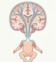
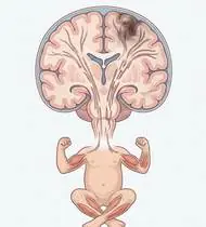
Cerebral Palsy (CP) is a group of permanent disorders of movement and posture causing activity limitation, attributed to non-progressive disturbances in the developing fetal or infant brain. Although the primary brain lesion (such as periventricular leukomalacia [PVL] in preterm infants causing spastic diplegia, or middle cerebral artery infarct causing spastic hemiplegia) is static, secondary musculoskeletal impairments (velocity-dependent spasticity, muscle shortening, bony torsional deformities, and spastic hip subluxation/dislocation) progress with growth! Classified functionally by GMFCS (Gross Motor Function Classification System Levels I–V) and MACS (Manual Ability Classification System Levels I–V).
Cerebral palsy (CP) is a condition caused by an early injury to the developing brain (before, during, or shortly after birth) that affects muscle tone, balance, and movement coordination. Crucially, the brain condition itself does not worsen, and because a child's brain has extraordinary neuroplasticity, play-based physiotherapy, slow stretching, and strengthening help children build lifelong motor skills and independence!
Primary Causes & Biomechanics
Key Points You Should Know
- •Prenatal, perinatal, or postnatal brain injury (prematurity <32 weeks / PVL, hypoxic-ischemic encephalopathy [HIE], perinatal stroke, or kernicterus)
- •Topographical & tonal subtypes: Spastic (~80%: Diplegia, Hemiplegia, Quadriplegia), Dyskinetic/Athetoid (~10–15%: basal ganglia lesion), and Ataxic (~5%: cerebellar lesion)
- •Velocity-Dependent Spasticity (Tardieu R1 vs. R2): Fast stretching triggers the R1 spastic catch; slow sustained stretching to R2 end-range and standing-frame weight-bearing preserve sarcomere length and acetabular hip centering!
- •Why Stretching Must Always Be Slow and Gentle: In spastic muscles, pulling the foot or leg *quickly* triggers an automatic reflex catch that makes the muscle tighten harder! Always stretch slowly, calmly, and playfully for 30 seconds.
- •Why 'W-Sitting' on the Floor Should Be Avoided: Sitting between the heels in a 'W' shape twists the thighbones inward and stresses the hip sockets—encourage criss-cross ('tailor') sitting, long sitting, or sitting on a small bench instead!
- •Strengthening Weak Muscles Is Safe and Essential: Modern research proves that strengthening a child's weak hip and thigh muscles (through bridges, squats, and stepping games) improves walking without making spasticity worse.
Assessment Methods
1. Goniometric ROM & Postural / Kinematic Analysis
GMFCS Level (I–V), GMFM-88 / GMFM-66 (Gross Motor Function Measure), Modified Tardieu Scale (measuring R1 fast spastic catch angle vs. R2 slow passive PROM!), and Modified Ashworth Scale (MAS).
2. Special Clinical Provocation & Neurological/Functional Tests
Hip Surveillance Protocol (Reimers Migration Percentage on AP Pelvis X-Ray — >30% indicates hip subluxation risk in GMFCS III–V!), Popliteal Angle (hamstrings), Silfverskiöld Test (gastrocnemius vs. soleus equinus), and primitive reflex persistence (ATNR, STNR, TLR, Moro).
3. Standardized Clinical Outcome Scales & Imaging Criteria
GMFCS (I–V), MACS (I–V), GMFM-66/88, Tardieu R1–R2 gap, and Reimers Hip Migration Percentage.
1. Range of Motion & Posture Check
Your physiotherapist gently evaluates how comfortably and smoothly your developing fetal or infant brain (periventricular white matter, motor cortex, basal ganglia, or cerebellum) and corticospinal pathways moves and checks your overall posture.
2. Muscle Strength & Functional Movement Check
Your muscle balance, walking or reaching comfort, and specific gentle movement tests are evaluated without triggering sharp pain.
3. Typical Symptoms & Daily Impact Review
Delayed Motor Milestones: Delay in head control (beyond 3–4 months), rolling, sitting independently (beyond 8–9 months), or walking (beyond 18 months).
Treatment Options & Phase-Based Protocol
✨ Enriched GIF Exercise & Precaution GuidePhase 1: Acute Symptom Modulation, Tissue Protection & Early Activation
- •Phase 1 Clinical Focus & Tissue Protection: Phase 1 — Slow Spasticity-Inhibiting Stretch, Handling & Postural Control Phase: Slow-velocity (never rapid ballistic!) gastro-soleus towel stretching, hamstring & hip adductor lengthening, Bobath / Neurodevelopmental Treatment (NDT) quadruped & midline trunk organization, and proper AFO / night positioning.
- •Pain, Edema & Neuromuscular Modulation: Targeted physical modalities, relative load management, and early protected mobility of the developing fetal or infant brain (periventricular white matter, motor cortex, basal ganglia, or cerebellum) and corticospinal pathways without provoking mechanical or inflammatory irritation.
- •Proximal & Distal Kinetic Chain Maintenance: Maintain active mobility and circulation in adjacent joints while protecting the primary healing structures in Cerebral Palsy (CP).
Phase 2: Progressive Mobility, Motor Control & Targeted Strengthening
- •Phase 2 Progressive Mobilization & Motor Control: Phase 2 — Antagonist & Functional Closed-Chain Strengthening Phase (Proven NOT to Increase Spasticity!): Supine pelvic bridging, side-lying/standing hip abduction, wall mini-squats & sit-to-stand transfers (targeting weak antigravity extensors and abductors to counter crouch and scissoring gait!).
- •Biomechanical Alignment & Co-Contraction: Restore balanced agonist-antagonist force couples, joint centration, and multi-planar neuromuscular control across the developing fetal or infant brain (periventricular white matter, motor cortex, basal ganglia, or cerebellum) and corticospinal pathways.
- •Myofascial Extensibility & Endurance: Address secondary compensatory tightness and build local muscular endurance within safe, symptom-free biomechanical parameters.
Phase 3: Functional Integration, Proprioception & Long-Term Prevention
- •Phase 3 Functional Integration & Long-Term Prevention: Phase 3 — Task-Specific Gait, Bimanual/CIMT & Hip Protection Phase: Supported tandem/balance play, Standing Frame in 30°–45° bilateral hip abduction (for GMFCS IV–V to drive femoral head into the acetabulum!), and CIMT / HABIT bimanual play in hemiplegic CP.
- •Proprioceptive Balance & Dynamic Loading: Progress to task-specific closed-chain, balance, and functional movement patterns tailored to daily life, occupational, or athletic demands.
- •Self-Management & Relapse Prophylaxis: Establish lifelong ergonomic habits, home exercise maintenance, and early warning-sign awareness to prevent recurrence.
Step 1: Calming Pain, Protecting the Area & Gentle Starter Motion
- •Why Stretching Must Always Be Slow and Gentle: In spastic muscles, pulling the foot or leg *quickly* triggers an automatic reflex catch that makes the muscle tighten harder! Always stretch slowly, calmly, and playfully for 30 seconds.
- •Calm Protection & Gentle Start: During the initial stage of Cerebral Palsy, avoid painful overloading and perform the gentle, pain-free starter movements shown below to support circulation and comfort.
- •Respecting Your Body's Signals: Every movement should stay within a comfortable, non-painful zone—never force through sharp or radiating pain.
Step 2: Restoring Flexibility & Building Supporting Muscle Strength
- •Why 'W-Sitting' on the Floor Should Be Avoided: Sitting between the heels in a 'W' shape twists the thighbones inward and stresses the hip sockets—encourage criss-cross ('tailor') sitting, long sitting, or sitting on a small bench instead!
- •Step-by-Step Flexibility & Support: As acute sensitivity settles, gradually build mobility and strengthen the supporting muscles around the joint at a steady, controlled pace.
- •Posture & Breathing Coordination: Keep your posture upright and breathe smoothly (never hold your breath!) during every repetition.
Step 3: Safe Daily Habits, Stamina & Preventing Recurrence
- •Strengthening Weak Muscles Is Safe and Essential: Modern research proves that strengthening a child's weak hip and thigh muscles (through bridges, squats, and stepping games) improves walking without making spasticity worse.
- •Confident Return to Daily Life: Integrate your stronger muscles and improved balance into walking, stairs, work, and recreational activities.
- •Preventing Future Flare-Ups: Continue your core protective habits and key maintenance exercises even after symptoms resolve.
Animated (GIF) Therapeutic Exercise Guide
Animated clinical exercise protocol with biomechanical angles, target muscle groups, and dosage parameters (Click image to enlarge)
Step-by-step animated exercise demonstrations you can safely perform at home (Click image to enlarge)
 🔍 Enlarge
1. Long-Sitting Towel-Assisted Gastrocnemius-Soleus & Plantar Fascia Stretch
1. Seated Towel Calf & Foot Arch Stretch
Long-sitting with a towel looped around the metatarsal heads, applying a slow, sustained passive pull into ankle dorsiflexion. Restores gastrocnemius-soleus-Achilles length and reduces plantar fascial and equine tension. Specifically indicated in Cerebral Palsy (CP) rehabilitation to optimize developing fetal or infant brain (periventricular white matter, motor cortex, basal ganglia, or cerebellum) and corticospinal pathways biomechanics and neuromuscular control.
Sit with your leg extended and loop a towel around the ball of your foot. Gently pull both ends of the towel toward you until you feel a comfortable stretch in your calf and sole, holding for 20–30 seconds. Perform smoothly within a comfortable, pain-free range to support your Cerebral Palsy recovery.
 🔍 Enlarge
2. Quadruped Contralateral Arm-Leg Reach ('Bird-Dog' Multifidus & Deep Core)
2. All-Fours Opposite Arm & Leg Reach (Bird-Dog)
In quadruped with a neutral lumbopelvic spine and abdominal drawing-in (transversus abdominis), extending opposite upper and lower extremities horizontally without pelvic rotation or lumbar hyperextension. Minimizes spinal compressive load while maximizing multifidus recruitment. Specifically indicated in Cerebral Palsy (CP) rehabilitation to optimize developing fetal or infant brain (periventricular white matter, motor cortex, basal ganglia, or cerebellum) and corticospinal pathways biomechanics and neuromuscular control.
On your hands and knees, keep your back flat like a tabletop and gently tighten your tummy. Slowly reach one arm forward and the opposite leg straight back withoutarching your lower back, hold for 5 seconds, and switch sides. Perform smoothly within a comfortable, pain-free range to support your Cerebral Palsy recovery.
 🔍 Enlarge
3. Side-Lying Hip Abduction (Gluteus Medius & Frontal Plane Control)
3. Side-Lying Leg Lift (Outer Hip Strengthening)
Isolated strengthening of the gluteus medius in slight hip extension and neutral rotation. Prevents contralateral pelvic drop (Trendelenburg) and reduces dynamic knee valgus and medial compartment adduction moments. Specifically indicated in Cerebral Palsy (CP) rehabilitation to optimize developing fetal or infant brain (periventricular white matter, motor cortex, basal ganglia, or cerebellum) and corticospinal pathways biomechanics and neuromuscular control.
Lie on your side with your bottom leg slightly bent for balance. Keep your top leg straight and slightly back, lift it up toward the ceiling without rolling your pelvis backward, hold for 3 seconds, and lower slowly. Perform smoothly within a comfortable, pain-free range to support your Cerebral Palsy recovery.
 🔍 Enlarge
4. Functional Closed-Kinetic-Chain Chair Transfer (Sit-to-Stand Biomechanics)
4. Controlled Chair Sit-to-Stand Transfer
Controlled transition from sitting to standing and eccentric lowering back to the chair with symmetrical weight distribution, anterior trunk inclination ('nose over toes'), and neutral frontal knee alignment. Specifically indicated in Cerebral Palsy (CP) rehabilitation to optimize developing fetal or infant brain (periventricular white matter, motor cortex, basal ganglia, or cerebellum) and corticospinal pathways biomechanics and neuromuscular control.
Sit near the front of a sturdy chair with your feet hip-width apart. Lean slightly forward and push evenly through both heels to stand up tall, then slowly lower yourself back down over 3 seconds. Perform smoothly within a comfortable, pain-free range to support your Cerebral Palsy recovery.
 🔍 Enlarge
5. Supine Lumbopelvic Bridging (Gluteus Maximus & Posterior Chain Co-Contraction)
5. Supine Hip Bridge (Controlled Pelvic Lift)
From hook-lying with transversus abdominis pre-activation, elevating the pelvis until hips, pelvis, and shoulders form a neutral diagonal line. Strengthens the gluteus maximus, hamstrings, and erector spinae synergy without lumbar hyperlordosis. Specifically indicated in Cerebral Palsy (CP) rehabilitation to optimize developing fetal or infant brain (periventricular white matter, motor cortex, basal ganglia, or cerebellum) and corticospinal pathways biomechanics and neuromuscular control.
Lie on your back with your knees bent and feet flat hip-width apart. Gently tighten your tummy and glutes, lift your hips until your body forms a straight line from shoulders to knees, hold for 5 seconds, and lower slowly. Perform smoothly within a comfortable, pain-free range to support your Cerebral Palsy recovery.
 🔍 Enlarge
6. Bobath Bilateral Clasp-Hand Elevation & Midline Trunk Organization
6. Clasped-Hands Two-Arm Overhead Reach
Interlocking the fingers with the affected thumb positioned on top, maintaining elbow extension and scapular protraction while elevating both arms symmetrically in the midline. Inhibits upper limb flexor synergy and prevents hemiplegic shoulder impingement. Specifically indicated in Cerebral Palsy (CP) rehabilitation to optimize developing fetal or infant brain (periventricular white matter, motor cortex, basal ganglia, or cerebellum) and corticospinal pathways biomechanics and neuromuscular control.
Clasp your hands together with your affected thumb on top. Keeping both elbows straight, use your stronger arm to smoothly guide both hands upward toward the ceiling and slowly back down. Perform smoothly within a comfortable, pain-free range to support your Cerebral Palsy recovery.
Movements to Avoid & Strict Precautions
1. Rapid, Jerky, or Forceful Stretching of Spastic Muscles (Triggers the Stretch Reflex Catch!)
1. Pulling the child's foot or arm quickly to stretch it (always move slowly and gently during play!)
2. Habitual 'W-Sitting' on the Floor (Increases Femoral Anteversion, In-Toeing & Hip Subluxation Risk!)
2. Letting the child sit in a 'W' position with feet tucked outside the hips (guide them into criss-cross or bench sitting)
3. Using Baby Walkers ('Jolly Jumpers') That Encourage Tiptoe Pushing & Scissoring!
3. Putting the child in wheeled baby-walkers or bouncers (worsens tiptoe stiffness and hip scissoring!)
4. Skipping Daily Standing-Frame Time or AFO Brace Wear During Growth Spurts
4. Leaving off prescribed ankle braces or standing-frame routines during rapid growth
Cerebral Palsy (CP) — Video Guide
Click directly on any video to open and watch in a window, or review details via 📋 Summary & Options.
Condition Overview Video (1 Video)
Summary of pathophysiology, biomechanics, and clinical presentation
What the condition is, why it develops, and its basic mechanism

What Is Cerebral Palsy (CP)? Subtypes and Clinical Characteristics
Cerebral Palsy (CP) is a group of permanent disorders of movement and posture causing activity limitation, attributed to non-progressive disturbances in the developing fetal or infant brain. Although the primary brain lesion (such as periventricular leukomalacia [PVL] in preterm infants causing spastic diplegia, or middle cerebral artery infarct causing spastic hemiplegia) is static, secondary musculoskeletal impairments (velocity-dependent spasticity, muscle shortening, bony torsional deformities, and spastic hip subluxation/dislocation) progress with growth! Classified functionally by GMFCS (Gross Motor Function Classification System Levels I–V) and MACS (Manual Ability Classification System Levels I–V).
Cerebral palsy (CP) is a condition caused by an early injury to the developing brain (before, during, or shortly after birth) that affects muscle tone, balance, and movement coordination. Crucially, the brain condition itself does not worsen, and because a child's brain has extraordinary neuroplasticity, play-based physiotherapy, slow stretching, and strengthening help children build lifelong motor skills and independence!
Treatment and Exercise Videos
Phase-based physiotherapy protocols, mobilization, and therapeutic exercises
Step-by-step recovery and safe home exercise applications

Home Exercises in Pediatric Neurological Conditions (CP, Spina Bifida, Brachial Plexus)
Phase 1: Acute Symptom Modulation, Tissue Protection & Early Activation: Phase 1 Clinical Focus & Tissue Protection: Phase 1 — Slow Spasticity-Inhibiting Stretch, Handling & Postural Control Phase: Slow-velocity (never rapid ballistic!) gastro-soleus towel stretching, hamstring & hip adductor lengthening, Bobath / Neurodevelopmental Treatment (NDT) quadruped & midline trunk organization, and proper AFO / night positioning.
Step 1: Calming Pain, Protecting the Area & Gentle Starter Motion: Why Stretching Must Always Be Slow and Gentle: In spastic muscles, pulling the foot or leg *quickly* triggers an automatic reflex catch that makes the muscle tighten harder! Always stretch slowly, calmly, and playfully for 30 seconds.

Trunk Control, Balance, and Functional Home Exercises in Pediatric Rehabilitation
Phase 2: Progressive Mobility, Motor Control & Targeted Strengthening: Phase 2 Progressive Mobilization & Motor Control: Phase 2 — Antagonist & Functional Closed-Chain Strengthening Phase (Proven NOT to Increase Spasticity!): Supine pelvic bridging, side-lying/standing hip abduction, wall mini-squats & sit-to-stand transfers (targeting weak antigravity extensors and abductors to counter crouch and scissoring gait!).
Step 2: Restoring Flexibility & Building Supporting Muscle Strength: Why 'W-Sitting' on the Floor Should Be Avoided: Sitting between the heels in a 'W' shape twists the thighbones inward and stresses the hip sockets—encourage criss-cross ('tailor') sitting, long sitting, or sitting on a small bench instead!
Clinical Assessment, Goniometry & Special Test Videos
Displayed only in Healthcare Professional mode (hidden in Patient mode)

Modified Ashworth Scale (MAS) — Clinical Spasticity Assessment
Goniometric ROM & Postural / Kinematic Analysis: GMFCS Level (I–V), GMFM-88 / GMFM-66 (Gross Motor Function Measure), Modified Tardieu Scale (measuring R1 fast spastic catch angle vs. R2 slow passive PROM!), and Modified Ashworth Scale (MAS).
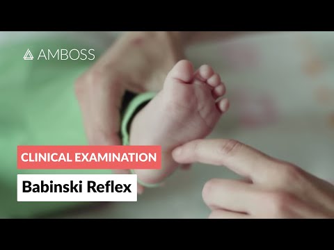
Pediatric Neurological Examination: Babinski and Plantar Reflex Assessment
Special Clinical Provocation & Neurological/Functional Tests: Hip Surveillance Protocol (Reimers Migration Percentage on AP Pelvis X-Ray — >30% indicates hip subluxation risk in GMFCS III–V!), Popliteal Angle (hamstrings), Silfverskiöld Test (gastrocnemius vs. soleus equinus), and primitive reflex persistence (ATNR, STNR, TLR, Moro).

Newborn and Infant Primitive Reflex Examination (Moro, Grasp, ATNR, Galant)
Standardized Clinical Outcome Scales & Imaging Criteria: GMFCS (I–V), MACS (I–V), GMFM-66/88, Tardieu R1–R2 gap, and Reimers Hip Migration Percentage.