Patellofemoral Pain Syndrome (Runner's / Moviegoe's Knee)Anterior Kneecap Pain (Runner's Knee) (Patellofemoral Pain Syndrome)
Patellofemoral Pain Syndrome (Runner's / Moviegoe's Knee) — Overview
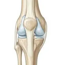
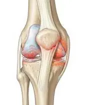
Patellofemoral Pain Syndrome (PFPS / 'Runner's Knee' or 'Movie-Goer's Sign') is a biomechanical overload of the retropatellar and peripatellar tissues driven by lateral patellar maltracking and dynamic knee valgus. Rather than isolated patellar pathology, modern biomechanics proves that femoral internal rotation and adduction beneath the patella (caused by proximal gluteus medius/maximus and hip external rotator weakness) decreases the patellofemoral contact area and spikes lateral facet joint reaction forces during stair descent, running, and squatting.
Patellofemoral pain syndrome (anterior kneecap pain / 'runner's knee') occurs when the kneecap (patella) pulls slightly toward the outer edge of its groove instead of gliding down the center—causing aching behind or around the kneecap during stairs, squatting, or sitting with bent knees.
Primary Causes & Biomechanics
Key Points You Should Know
- •Proximal factor: Hip abductor and external rotator weakness allowing dynamic knee valgus (femoral adduction + internal rotation) during single-leg stance
- •Local factor: Delayed or weakened Vastus Medialis Obliquus (VMO) activation relative to Vastus Lateralis (VL) and tight lateral retinaculum / iliotibial band (ITB)
- •Distal factor: Excessive subtalar foot pronation causing compensatory internal tibial/femoral rotation
- •Why Hip Strength Fixes Kneecap Pain: The groove your kneecap sits in is part of your thighbone; when your outer hip muscles are strong, they stop your thighbone from rotating inward under the kneecap!
- •The 'Movie-Goer's Sign': Sitting for a long time with bent knees presses the kneecap tightly against the thighbone—briefly straightening your legs relieves that pressure.
- •Keep Knees Over Second Toes: Whenever you squat or use stairs, never let your knees cave inward toward each other.
Assessment Methods
1. Goniometric ROM & Postural / Kinematic Analysis
Knee Q-angle / dynamic Frontal Plane Projection Angle (FPPA) during a single-leg step-down, patellar glide/tilt mobility, and Ober's test (ITB tightness) / Ely's test (rectus femoris).
2. Special Clinical Provocation & Neurological/Functional Tests
Patellar Grind (Clarke's) Test, Waldron's Squat Test, Eccentric Step-Down Test (most reliable clinical functional test!), and McConnell medial patellar glide symptom-modification test.
3. Standardized Clinical Outcome Scales & Imaging Criteria
Kujala Anterior Knee Pain Scale (AKPS), KOOS-PF, and Merchant/Skyline patellar radiographs.
1. Range of Motion & Posture Check
Your physiotherapist gently evaluates how comfortably and smoothly your patellofemoral joint (retropatellar hyaline cartilage, femoral trochlear groove, medial/lateral retinaculum, and VMO/gluteal stabilizers) moves and checks your overall posture.
2. Muscle Strength & Functional Movement Check
Your muscle balance, walking or reaching comfort, and specific gentle movement tests are evaluated without triggering sharp pain.
3. Typical Symptoms & Daily Impact Review
Diffuse, aching pain behind, underneath, or around the kneecap (retropatellar/peripatellar) aggravated by walking DOWN stairs, squatting, lunging, running (especially downhill), or sitting for long periods with bent knees ('Movie-Goer's / Theater Sign').
Treatment Options & Phase-Based Protocol
✨ Enriched GIF Exercise & Precaution GuidePhase 1: Acute Symptom Modulation, Tissue Protection & Early Activation
- •Phase 1 Clinical Focus & Tissue Protection: Pain Relief, Patellar Centering & Isometric VMO Phase (Weeks 1–2): Load modification, McConnell / Kinesio medial patellar taping, isometric quadriceps/VMO setting at 0° extension (where patellofemoral joint reaction force is ~0!), and 4-way straight leg raises (SLR).
- •Pain, Edema & Neuromuscular Modulation: Targeted physical modalities, relative load management, and early protected mobility of the patellofemoral joint (retropatellar hyaline cartilage, femoral trochlear groove, medial/lateral retinaculum, and VMO/gluteal stabilizers) without provoking mechanical or inflammatory irritation.
- •Proximal & Distal Kinetic Chain Maintenance: Maintain active mobility and circulation in adjacent joints while protecting the primary healing structures in Patellofemoral Pain Syndrome.
Phase 2: Progressive Mobility, Motor Control & Targeted Strengthening
- •Phase 2 Progressive Mobilization & Motor Control: Proximal Hip + Safe-Angle Closed-Chain Phase (Weeks 2–6): Combined Hip + Knee rehabilitation (proven superior to knee-only rehab!)—side-lying & standing hip abduction, supine pelvic bridging, seated terminal knee extension (30°–0°), and protected 0°–40° wall mini-squats.
- •Biomechanical Alignment & Co-Contraction: Restore balanced agonist-antagonist force couples, joint centration, and multi-planar neuromuscular control across the patellofemoral joint (retropatellar hyaline cartilage, femoral trochlear groove, medial/lateral retinaculum, and VMO/gluteal stabilizers).
- •Myofascial Extensibility & Endurance: Address secondary compensatory tightness and build local muscular endurance within safe, symptom-free biomechanical parameters.
Phase 3: Functional Integration, Proprioception & Long-Term Prevention
- •Phase 3 Functional Integration & Long-Term Prevention: Dynamic Alignment & Eccentric Control Phase (Week 6–12): Mirror-guided chair sit-to-stand and low step-downs (keeping the kneecap aligned over the 2nd toe without caving inward!), towel calf/hamstring/quad stretching, and running cadence retraining (+5–10% step rate reduces PFJ load by 15–20%).
- •Proprioceptive Balance & Dynamic Loading: Progress to task-specific closed-chain, balance, and functional movement patterns tailored to daily life, occupational, or athletic demands.
- •Self-Management & Relapse Prophylaxis: Establish lifelong ergonomic habits, home exercise maintenance, and early warning-sign awareness to prevent recurrence.
Step 1: Calming Pain, Protecting the Area & Gentle Starter Motion
- •Why Hip Strength Fixes Kneecap Pain: The groove your kneecap sits in is part of your thighbone; when your outer hip muscles are strong, they stop your thighbone from rotating inward under the kneecap!
- •Calm Protection & Gentle Start: During the initial stage of Anterior Kneecap Pain (Runner's Knee), avoid painful overloading and perform the gentle, pain-free starter movements shown below to support circulation and comfort.
- •Respecting Your Body's Signals: Every movement should stay within a comfortable, non-painful zone—never force through sharp or radiating pain.
Step 2: Restoring Flexibility & Building Supporting Muscle Strength
- •The 'Movie-Goer's Sign': Sitting for a long time with bent knees presses the kneecap tightly against the thighbone—briefly straightening your legs relieves that pressure.
- •Step-by-Step Flexibility & Support: As acute sensitivity settles, gradually build mobility and strengthen the supporting muscles around the joint at a steady, controlled pace.
- •Posture & Breathing Coordination: Keep your posture upright and breathe smoothly (never hold your breath!) during every repetition.
Step 3: Safe Daily Habits, Stamina & Preventing Recurrence
- •Keep Knees Over Second Toes: Whenever you squat or use stairs, never let your knees cave inward toward each other.
- •Confident Return to Daily Life: Integrate your stronger muscles and improved balance into walking, stairs, work, and recreational activities.
- •Preventing Future Flare-Ups: Continue your core protective habits and key maintenance exercises even after symptoms resolve.
Animated (GIF) Therapeutic Exercise Guide
Animated clinical exercise protocol with biomechanical angles, target muscle groups, and dosage parameters (Click image to enlarge)
Step-by-step animated exercise demonstrations you can safely perform at home (Click image to enlarge)
 🔍 Enlarge
1. Isometric Quadriceps Setting (Popliteal Towel Press)
1. Under-Knee Towel Press (Quadriceps Tightening)
Isometric compression of a rolled towel beneath the popliteal fossa into the surface with simultaneous ankle dorsiflexion. Activates the Vastus Medialis Obliquus (VMO) and overcomes arthrogenic muscle inhibition with 0° joint shear stress. Specifically indicated in Patellofemoral Pain Syndrome rehabilitation to optimize patellofemoral joint (retropatellar hyaline cartilage, femoral trochlear groove, medial/lateral retinaculum, and VMO/gluteal stabilizers) biomechanics and neuromuscular control.
Place a small rolled towel under your knee while lying on your back or sitting with your leg straight. Pull your toes toward you while firmly pressing the back of your knee down into the towel, hold for 5 seconds, and relax while exhaling. Perform smoothly within a comfortable, pain-free range to support your Anterior Kneecap Pain (Runner's Knee) recovery.
 🔍 Enlarge
2. Straight Leg Raise (SLR — Multi-Plane Isometric Knee Lock)
2. Supine Straight Leg Raise
Maintaining the knee locked in full extension (0°) with ankle dorsiflexion while elevating the lower extremity to 45° hip flexion. Strengthens the quadriceps and iliopsoas complex without compressing or shearing the articular surfaces. Specifically indicated in Patellofemoral Pain Syndrome rehabilitation to optimize patellofemoral joint (retropatellar hyaline cartilage, femoral trochlear groove, medial/lateral retinaculum, and VMO/gluteal stabilizers) biomechanics and neuromuscular control.
Bend your healthy knee with the foot flat on the bed. Tighten the thigh muscle of your affected leg to lock the knee straight, lift the leg to the height of the opposite knee, hold for 3 seconds, and lower slowly. Perform smoothly within a comfortable, pain-free range to support your Anterior Kneecap Pain (Runner's Knee) recovery.
 🔍 Enlarge
3. Side-Lying Hip Abduction (Gluteus Medius & Frontal Plane Control)
3. Side-Lying Leg Lift (Outer Hip Strengthening)
Isolated strengthening of the gluteus medius in slight hip extension and neutral rotation. Prevents contralateral pelvic drop (Trendelenburg) and reduces dynamic knee valgus and medial compartment adduction moments. Specifically indicated in Patellofemoral Pain Syndrome rehabilitation to optimize patellofemoral joint (retropatellar hyaline cartilage, femoral trochlear groove, medial/lateral retinaculum, and VMO/gluteal stabilizers) biomechanics and neuromuscular control.
Lie on your side with your bottom leg slightly bent for balance. Keep your top leg straight and slightly back, lift it up toward the ceiling without rolling your pelvis backward, hold for 3 seconds, and lower slowly. Perform smoothly within a comfortable, pain-free range to support your Anterior Kneecap Pain (Runner's Knee) recovery.
 🔍 Enlarge
4. Protected Closed-Kinetic-Chain Wall Mini-Squat (0°–40° Safe Arc)
4. Wall-Supported Safe Mini-Squat
Closed-kinetic-chain bilateral knee flexion in the safe 0°–40° arc with the back supported against a wall. Promotes quadriceps-hamstring-gluteal co-contraction and joint proprioception without excessive patellofemoral compression. Specifically indicated in Patellofemoral Pain Syndrome rehabilitation to optimize patellofemoral joint (retropatellar hyaline cartilage, femoral trochlear groove, medial/lateral retinaculum, and VMO/gluteal stabilizers) biomechanics and neuromuscular control.
Lean your back flat against a wall with your feet shoulder-width apart. Slowly bend your knees slightly (30–40 degrees, keeping knees behind toes), hold for 5 seconds, and push back up. Perform smoothly within a comfortable, pain-free range to support your Anterior Kneecap Pain (Runner's Knee) recovery.
 🔍 Enlarge
5. Seated Terminal Knee Extension (Open/Closed Chain VMO Focus)
5. Seated Knee Straightening & Hold
Concentric extension of the knee to the final 30°–0° terminal arc with isometric hold and ankle dorsiflexion. Restores terminal knee extension torque and stabilizes dynamic patellofemoral tracking. Specifically indicated in Patellofemoral Pain Syndrome rehabilitation to optimize patellofemoral joint (retropatellar hyaline cartilage, femoral trochlear groove, medial/lateral retinaculum, and VMO/gluteal stabilizers) biomechanics and neuromuscular control.
Sit upright in a chair and slowly straighten your knee completely while pulling your toes toward you. Feel the front thigh muscle tighten, hold for 5 seconds, and slowly lower your foot. Perform smoothly within a comfortable, pain-free range to support your Anterior Kneecap Pain (Runner's Knee) recovery.
 🔍 Enlarge
6. Long-Sitting Towel-Assisted Gastrocnemius-Soleus & Plantar Fascia Stretch
6. Seated Towel Calf & Foot Arch Stretch
Long-sitting with a towel looped around the metatarsal heads, applying a slow, sustained passive pull into ankle dorsiflexion. Restores gastrocnemius-soleus-Achilles length and reduces plantar fascial and equine tension. Specifically indicated in Patellofemoral Pain Syndrome rehabilitation to optimize patellofemoral joint (retropatellar hyaline cartilage, femoral trochlear groove, medial/lateral retinaculum, and VMO/gluteal stabilizers) biomechanics and neuromuscular control.
Sit with your leg extended and loop a towel around the ball of your foot. Gently pull both ends of the towel toward you until you feel a comfortable stretch in your calf and sole, holding for 20–30 seconds. Perform smoothly within a comfortable, pain-free range to support your Anterior Kneecap Pain (Runner's Knee) recovery.
Movements to Avoid & Strict Precautions
1. Deep Squats (>60–90°), Deep Lunges, & Allowing the Knees to Cave Inward (Valgus)
1. Deep squats, deep lunges, or letting your knees knock inward when squatting or descending stairs
2. Heavy Seated Leg-Extension Machine Lifts Through Full 90°–0° Arc
2. Using heavy seated leg-extension machines at the gym (grinds the kneecap against the groove!)
3. Sitting for Hours With Knees Tightly Tucked Under the Chair
3. Sitting for hours with your knees bent tightly underneath your chair
4. Running Down Steep Hills or With a Long Over-Striding Heel Strike
4. Running down steep hills or taking long bounding strides when running
Patellofemoral Pain Syndrome (Runner's / Moviegoe's Knee) — Video Guide
Click directly on any video to open and watch in a window, or review details via 📋 Summary & Options.
Condition Overview Video (1 Video)
Summary of pathophysiology, biomechanics, and clinical presentation
What the condition is, why it develops, and its basic mechanism

What Is Patellofemoral Pain Syndrome (Chondromalacia)? How Is It Treated?
Patellofemoral Pain Syndrome (PFPS / 'Runner's Knee' or 'Movie-Goer's Sign') is a biomechanical overload of the retropatellar and peripatellar tissues driven by lateral patellar maltracking and dynamic knee valgus. Rather than isolated patellar pathology, modern biomechanics proves that femoral internal rotation and adduction beneath the patella (caused by proximal gluteus medius/maximus and hip external rotator weakness) decreases the patellofemoral contact area and spikes lateral facet joint reaction forces during stair descent, running, and squatting.
Patellofemoral pain syndrome (anterior kneecap pain / 'runner's knee') occurs when the kneecap (patella) pulls slightly toward the outer edge of its groove instead of gliding down the center—causing aching behind or around the kneecap during stairs, squatting, or sitting with bent knees.
Treatment and Exercise Videos
Phase-based physiotherapy protocols, mobilization, and therapeutic exercises
Step-by-step recovery and safe home exercise applications

Anterior Knee Pain and Patellofemoral Pain Syndrome Physiotherapy Exercises
Phase 1: Acute Symptom Modulation, Tissue Protection & Early Activation: Phase 1 Clinical Focus & Tissue Protection: Pain Relief, Patellar Centering & Isometric VMO Phase (Weeks 1–2): Load modification, McConnell / Kinesio medial patellar taping, isometric quadriceps/VMO setting at 0° extension (where patellofemoral joint reaction force is ~0!), and 4-way straight leg raises (SLR).
Step 1: Calming Pain, Protecting the Area & Gentle Starter Motion: Why Hip Strength Fixes Kneecap Pain: The groove your kneecap sits in is part of your thighbone; when your outer hip muscles are strong, they stop your thighbone from rotating inward under the kneecap!

What Is Patellofemoral Pain Syndrome? 4 Essential Home Exercises
Phase 2: Progressive Mobility, Motor Control & Targeted Strengthening: Phase 2 Progressive Mobilization & Motor Control: Proximal Hip + Safe-Angle Closed-Chain Phase (Weeks 2–6): Combined Hip + Knee rehabilitation (proven superior to knee-only rehab!)—side-lying & standing hip abduction, supine pelvic bridging, seated terminal knee extension (30°–0°), and protected 0°–40° wall mini-squats.
Step 2: Restoring Flexibility & Building Supporting Muscle Strength: The 'Movie-Goer's Sign': Sitting for a long time with bent knees presses the kneecap tightly against the thighbone—briefly straightening your legs relieves that pressure.
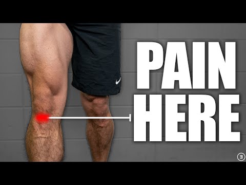
Strengthening Exercises for Patellofemoral Pain Syndrome and Anterior Knee Pain
Phase 3: Functional Integration, Proprioception & Long-Term Prevention: Phase 3 Functional Integration & Long-Term Prevention: Dynamic Alignment & Eccentric Control Phase (Week 6–12): Mirror-guided chair sit-to-stand and low step-downs (keeping the kneecap aligned over the 2nd toe without caving inward!), towel calf/hamstring/quad stretching, and running cadence retraining (+5–10% step rate reduces PFJ load by 15–20%).
Step 3: Safe Daily Habits, Stamina & Preventing Recurrence: Keep Knees Over Second Toes: Whenever you squat or use stairs, never let your knees cave inward toward each other.
Clinical Assessment, Goniometry & Special Test Videos
Displayed only in Healthcare Professional mode (hidden in Patient mode)

Patellar Grind (Clarke's) Test — Patellofemoral Cartilage and Crepitus Examination
Goniometric ROM & Postural / Kinematic Analysis: Knee Q-angle / dynamic Frontal Plane Projection Angle (FPPA) during a single-leg step-down, patellar glide/tilt mobility, and Ober's test (ITB tightness) / Ely's test (rectus femoris).

Systematic Knee Joint Physical Examination and Clinical Assessment
Special Clinical Provocation & Neurological/Functional Tests: Patellar Grind (Clarke's) Test, Waldron's Squat Test, Eccentric Step-Down Test (most reliable clinical functional test!), and McConnell medial patellar glide symptom-modification test.

Knee Joint Goniometric Measurement (Active & Passive Flexion / Extension ROM)
Standardized Clinical Outcome Scales & Imaging Criteria: Kujala Anterior Knee Pain Scale (AKPS), KOOS-PF, and Merchant/Skyline patellar radiographs.