Lumbar Spondylosis (Degenerative Lumbar Spine)Lower Back Arthritis (Wear & Tear) (Lumbar Spondylosis)
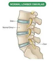
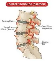
Lumbar Spondylosis (Degenerative Lumbar Spine) — Overview
Lumbar spondylosis is a chronic degenerative cascade of the lumbar motion segment starting with intervertebral disc dehydration and height loss, which transfers up to 40–70% of axial compressive load onto the posterior zygapophyseal (facet) joints, resulting in facet arthrosis, marginal osteophytes, and capsular hypertrophy.
Lumbar spondylosis (lower back osteoarthritis / facet wear-and-tear) is the natural age-related thinning of the spinal disc cushions and roughening of the small stabilizing joints (facet joints) at the back of the lumbar spine.
Primary Causes & Biomechanics
Key Points You Should Know
- •Age-related proteoglycan loss in the nucleus pulposus and repetitive mechanical axial/rotational loading
- •Increased lumbar hyperlordosis and anterior pelvic tilt compressing the posterior facet joint surfaces
- •Weakness of the abdominal core (transversus abdominis/obliques) and tightness of the iliopsoas/hamstrings
- •Morning Stiffness That Warms Up: Just like a rusty hinge, spondylosis feels stiff for the first 15–30 minutes in the morning and loosens up once you apply warmth and gentle movement.
- •Posterior Pelvic Tilt Relief: Gently flattening your excessive lower back arch relieves pinching on the rear spinal joints.
- •Active Core Support: Strong tummy and hip muscles act like shock absorbers for your lower back.
Assessment Methods
1. Goniometric ROM & Postural / Kinematic Analysis
Lumbar flexion/extension/lateral flexion ROM (extension and quadrant rotation typically provoke facet pain; Modified Schober test).
2. Special Clinical Provocation & Neurological/Functional Tests
Kemp's (Lumbar Quadrant Extension-Rotation) Test for facet joint provocation, spinous/transverse process springing (PA glide), and absence of acute nerve root tension signs.
3. Standardized Clinical Outcome Scales & Imaging Criteria
Oswestry Disability Index (ODI), Roland-Morris Questionnaire, and standing lumbar radiographs.
1. Range of Motion & Posture Check
Your physiotherapist gently evaluates how comfortably and smoothly your lumbar intervertebral discs, vertebral endplates, and zygapophyseal (facet) joints moves and checks your overall posture.
2. Muscle Strength & Functional Movement Check
Your muscle balance, walking or reaching comfort, and specific gentle movement tests are evaluated without triggering sharp pain.
3. Typical Symptoms & Daily Impact Review
Aching lower back pain and morning stiffness (<30 min) that improves with gentle movement, worsens with prolonged standing or arching backward, and may refer dull ache to the buttocks/posterior thighs (above the knee).
Treatment Options & Phase-Based Protocol
✨ Enriched GIF Exercise & Precaution GuidePhase 1: Acute Symptom Modulation, Tissue Protection & Early Activation
- •Phase 1 Clinical Focus & Tissue Protection: Pain Relief, Thermal Therapy & Unloading Phase (Weeks 1–2): Superficial/deep heat + TENS for morning stiffness, supine posterior pelvic tilts, and unloaded supine heel slides.
- •Pain, Edema & Neuromuscular Modulation: Targeted physical modalities, relative load management, and early protected mobility of the lumbar intervertebral discs, vertebral endplates, and zygapophyseal (facet) joints without provoking mechanical or inflammatory irritation.
- •Proximal & Distal Kinetic Chain Maintenance: Maintain active mobility and circulation in adjacent joints while protecting the primary healing structures in Lumbar Spondylosis.
Phase 2: Progressive Mobility, Motor Control & Targeted Strengthening
- •Phase 2 Progressive Mobilization & Motor Control: Lumbopelvic Core & Facet Decompression Phase (Weeks 2–6): Supine pelvic bridging, quadruped Bird-Dog stabilization, and piriformis/gluteal/hamstring stretching to reduce anterior pelvic tilt and facet compression.
- •Biomechanical Alignment & Co-Contraction: Restore balanced agonist-antagonist force couples, joint centration, and multi-planar neuromuscular control across the lumbar intervertebral discs, vertebral endplates, and zygapophyseal (facet) joints.
- •Myofascial Extensibility & Endurance: Address secondary compensatory tightness and build local muscular endurance within safe, symptom-free biomechanical parameters.
Phase 3: Functional Integration, Proprioception & Long-Term Prevention
- •Phase 3 Functional Integration & Long-Term Prevention: Aerobic Conditioning & Postural Endurance Phase (Week 6+): Wall-referenced axial extension, controlled chair sit-to-stand, daily walking or aquatic therapy, and ergonomic lumbar support.
- •Proprioceptive Balance & Dynamic Loading: Progress to task-specific closed-chain, balance, and functional movement patterns tailored to daily life, occupational, or athletic demands.
- •Self-Management & Relapse Prophylaxis: Establish lifelong ergonomic habits, home exercise maintenance, and early warning-sign awareness to prevent recurrence.
Step 1: Calming Pain, Protecting the Area & Gentle Starter Motion
- •Morning Stiffness That Warms Up: Just like a rusty hinge, spondylosis feels stiff for the first 15–30 minutes in the morning and loosens up once you apply warmth and gentle movement.
- •Calm Protection & Gentle Start: During the initial stage of Lower Back Arthritis (Wear & Tear), avoid painful overloading and perform the gentle, pain-free starter movements shown below to support circulation and comfort.
- •Respecting Your Body's Signals: Every movement should stay within a comfortable, non-painful zone—never force through sharp or radiating pain.
Step 2: Restoring Flexibility & Building Supporting Muscle Strength
- •Posterior Pelvic Tilt Relief: Gently flattening your excessive lower back arch relieves pinching on the rear spinal joints.
- •Step-by-Step Flexibility & Support: As acute sensitivity settles, gradually build mobility and strengthen the supporting muscles around the joint at a steady, controlled pace.
- •Posture & Breathing Coordination: Keep your posture upright and breathe smoothly (never hold your breath!) during every repetition.
Step 3: Safe Daily Habits, Stamina & Preventing Recurrence
- •Active Core Support: Strong tummy and hip muscles act like shock absorbers for your lower back.
- •Confident Return to Daily Life: Integrate your stronger muscles and improved balance into walking, stairs, work, and recreational activities.
- •Preventing Future Flare-Ups: Continue your core protective habits and key maintenance exercises even after symptoms resolve.
Animated (GIF) Therapeutic Exercise Guide
Animated clinical exercise protocol with biomechanical angles, target muscle groups, and dosage parameters (Click image to enlarge)
Step-by-step animated exercise demonstrations you can safely perform at home (Click image to enlarge)
 🔍 Enlarge
1. Supine Active-Assisted Heel Slide & Synovial Diffusion Mobilization
1. Supine Heel Slide (Gentle Knee & Hip Bending)
Controlled flexion and extension of the lower extremity by sliding the heel along the support surface without axial weight-bearing. Stimulates synovial fluid diffusion across avascular cartilage and prevents capsular adhesions. Specifically indicated in Lumbar Spondylosis rehabilitation to optimize lumbar intervertebral discs, vertebral endplates, and zygapophyseal (facet) joints biomechanics and neuromuscular control.
While lying on your back, slowly slide your heel along the bed sheet toward your hip within a comfortable, pain-free angle, hold for 3 seconds, and gently slide it back out straight. Perform smoothly within a comfortable, pain-free range to support your Lower Back Arthritis (Wear & Tear) recovery.
 🔍 Enlarge
2. Quadruped Contralateral Arm-Leg Reach ('Bird-Dog' Multifidus & Deep Core)
2. All-Fours Opposite Arm & Leg Reach (Bird-Dog)
In quadruped with a neutral lumbopelvic spine and abdominal drawing-in (transversus abdominis), extending opposite upper and lower extremities horizontally without pelvic rotation or lumbar hyperextension. Minimizes spinal compressive load while maximizing multifidus recruitment. Specifically indicated in Lumbar Spondylosis rehabilitation to optimize lumbar intervertebral discs, vertebral endplates, and zygapophyseal (facet) joints biomechanics and neuromuscular control.
On your hands and knees, keep your back flat like a tabletop and gently tighten your tummy. Slowly reach one arm forward and the opposite leg straight back withoutarching your lower back, hold for 5 seconds, and switch sides. Perform smoothly within a comfortable, pain-free range to support your Lower Back Arthritis (Wear & Tear) recovery.
 🔍 Enlarge
3. Side-Lying Hip Abduction (Gluteus Medius & Frontal Plane Control)
3. Side-Lying Leg Lift (Outer Hip Strengthening)
Isolated strengthening of the gluteus medius in slight hip extension and neutral rotation. Prevents contralateral pelvic drop (Trendelenburg) and reduces dynamic knee valgus and medial compartment adduction moments. Specifically indicated in Lumbar Spondylosis rehabilitation to optimize lumbar intervertebral discs, vertebral endplates, and zygapophyseal (facet) joints biomechanics and neuromuscular control.
Lie on your side with your bottom leg slightly bent for balance. Keep your top leg straight and slightly back, lift it up toward the ceiling without rolling your pelvis backward, hold for 3 seconds, and lower slowly. Perform smoothly within a comfortable, pain-free range to support your Lower Back Arthritis (Wear & Tear) recovery.
 🔍 Enlarge
4. Functional Closed-Kinetic-Chain Chair Transfer (Sit-to-Stand Biomechanics)
4. Controlled Chair Sit-to-Stand Transfer
Controlled transition from sitting to standing and eccentric lowering back to the chair with symmetrical weight distribution, anterior trunk inclination ('nose over toes'), and neutral frontal knee alignment. Specifically indicated in Lumbar Spondylosis rehabilitation to optimize lumbar intervertebral discs, vertebral endplates, and zygapophyseal (facet) joints biomechanics and neuromuscular control.
Sit near the front of a sturdy chair with your feet hip-width apart. Lean slightly forward and push evenly through both heels to stand up tall, then slowly lower yourself back down over 3 seconds. Perform smoothly within a comfortable, pain-free range to support your Lower Back Arthritis (Wear & Tear) recovery.
 🔍 Enlarge
5. Supine Posterior Pelvic Tilt & Transversus Abdominis Activation (Abdominal Hollowing)
5. Supine Lower Back Flatten (Pelvic Tilt & Deep Core)
In hook-lying, gently drawing the umbilicus inward-upward (transversus abdominis) while tilting the pelvis posteriorly to flatten excessive lumbar lordosis against the mat. Decompresses posterior facet joints and narrows the intervertebral shear vector. Specifically indicated in Lumbar Spondylosis rehabilitation to optimize lumbar intervertebral discs, vertebral endplates, and zygapophyseal (facet) joints biomechanics and neuromuscular control.
Lie on your back with your knees bent. Without holding your breath, gently draw your belly button inward and press the small of your back flat against the bed, hold for 5 seconds, and relax. Perform smoothly within a comfortable, pain-free range to support your Lower Back Arthritis (Wear & Tear) recovery.
 🔍 Enlarge
6. Supine Lumbopelvic Bridging (Gluteus Maximus & Posterior Chain Co-Contraction)
6. Supine Hip Bridge (Controlled Pelvic Lift)
From hook-lying with transversus abdominis pre-activation, elevating the pelvis until hips, pelvis, and shoulders form a neutral diagonal line. Strengthens the gluteus maximus, hamstrings, and erector spinae synergy without lumbar hyperlordosis. Specifically indicated in Lumbar Spondylosis rehabilitation to optimize lumbar intervertebral discs, vertebral endplates, and zygapophyseal (facet) joints biomechanics and neuromuscular control.
Lie on your back with your knees bent and feet flat hip-width apart. Gently tighten your tummy and glutes, lift your hips until your body forms a straight line from shoulders to knees, hold for 5 seconds, and lower slowly. Perform smoothly within a comfortable, pain-free range to support your Lower Back Arthritis (Wear & Tear) recovery.
Movements to Avoid & Strict Precautions
1. Sustained Lumbar Hyperextension (Arching Backward) or Prolonged Standing in One Spot
1. Standing still for hours with a swayed back or arching backward forcefully
2. Heavy Bending and Twisting Without Hip-Hinge Mechanics
2. Lifting heavy loads with a twisted or rounded back
3. Sleeping on a Sagging Mattress or on Your Stomach (Increases Facet Pinching!)
3. Sleeping on your stomach, which arches and pinches the lower back joints
4. Complete Sedentary Inactivity (Accelerates Joint Stiffness and Muscle Weakness)
4. Staying completely inactive, which makes spinal joints stiffer
Lumbar Spondylosis (Degenerative Lumbar Spine) — Video Guide
Click directly on any video to open and watch in a window, or review details via 📋 Summary & Options.
Condition Overview Video (1 Video)
Summary of pathophysiology, biomechanics, and clinical presentation
What the condition is, why it develops, and its basic mechanism

Clinical Differential Diagnosis of Lumbar Spondylosis, Spinal Stenosis, and Spondylolisthesis
Lumbar spondylosis is a chronic degenerative cascade of the lumbar motion segment starting with intervertebral disc dehydration and height loss, which transfers up to 40–70% of axial compressive load onto the posterior zygapophyseal (facet) joints, resulting in facet arthrosis, marginal osteophytes, and capsular hypertrophy.
Lumbar spondylosis (lower back osteoarthritis / facet wear-and-tear) is the natural age-related thinning of the spinal disc cushions and roughening of the small stabilizing joints (facet joints) at the back of the lumbar spine.
Treatment and Exercise Videos
Phase-based physiotherapy protocols, mobilization, and therapeutic exercises
Step-by-step recovery and safe home exercise applications
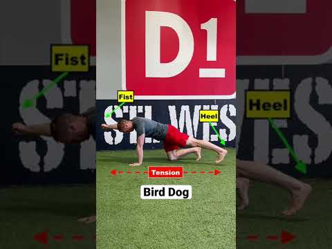
Lumbar Disc Herniation Exercises | Progressive Physiotherapy and Core Stabilization
Phase 1: Acute Symptom Modulation, Tissue Protection & Early Activation: Phase 1 Clinical Focus & Tissue Protection: Pain Relief, Thermal Therapy & Unloading Phase (Weeks 1–2): Superficial/deep heat + TENS for morning stiffness, supine posterior pelvic tilts, and unloaded supine heel slides.
Step 1: Calming Pain, Protecting the Area & Gentle Starter Motion: Morning Stiffness That Warms Up: Just like a rusty hinge, spondylosis feels stiff for the first 15–30 minutes in the morning and loosens up once you apply warmth and gentle movement.

What Is Lumbar Spinal Stenosis? Flexion-Based Exercise Therapy Explained
Phase 2: Progressive Mobility, Motor Control & Targeted Strengthening: Phase 2 Progressive Mobilization & Motor Control: Lumbopelvic Core & Facet Decompression Phase (Weeks 2–6): Supine pelvic bridging, quadruped Bird-Dog stabilization, and piriformis/gluteal/hamstring stretching to reduce anterior pelvic tilt and facet compression.
Step 2: Restoring Flexibility & Building Supporting Muscle Strength: Posterior Pelvic Tilt Relief: Gently flattening your excessive lower back arch relieves pinching on the rear spinal joints.
Clinical Assessment, Goniometry & Special Test Videos
Displayed only in Healthcare Professional mode (hidden in Patient mode)

Lumbar Spine Range of Motion and Clinical Physical Examination
Goniometric ROM & Postural / Kinematic Analysis: Lumbar flexion/extension/lateral flexion ROM (extension and quadrant rotation typically provoke facet pain; Modified Schober test).
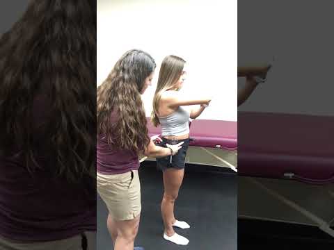
Vertebral Column (Cervical & Thoracolumbar Spine) Goniometric and Tape Measurement
Special Clinical Provocation & Neurological/Functional Tests: Kemp's (Lumbar Quadrant Extension-Rotation) Test for facet joint provocation, spinous/transverse process springing (PA glide), and absence of acute nerve root tension signs.