Gonarthrosis (Knee Osteoarthritis)Knee Osteoarthritis (Wear & Tear) (Gonarthrosis (Knee Osteoarthritis))
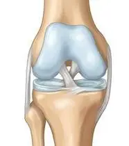
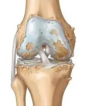
Gonarthrosis (Knee Osteoarthritis) — Overview
Gonarthrosis (knee osteoarthritis) is a chronic degenerative joint disorder characterized by progressive loss of articular hyaline cartilage, subchondral bone sclerosis, marginal osteophyte formation, and synovial inflammation, affecting load transmission across the medial, lateral, and patellofemoral compartments.
Gonarthrosis (commonly known as knee osteoarthritis or wear-and-tear arthritis) is the gradual thinning and roughening of the smooth cartilage cushion covering the ends of the bones inside your knee joint. As this natural shock absorber wears down, the knee experiences friction, brief morning stiffness, and aching during weight-bearing.
Primary Causes & Biomechanics
Key Points You Should Know
- •Frontal plane malalignment (particularly genu varum) increasing the external knee adduction moment (KAM) across the medial compartment
- •Advanced age, elevated BMI, and history of prior meniscal tear or anterior cruciate ligament (ACL) injury
- •Arthrogenic muscle inhibition (AMI) and weakness of the quadriceps (VMO) and hip abductor (gluteus medius) musculature
- •Articular Cartilage Cushion: The smooth, slippery coating that allows knee bones to glide effortlessly without rubbing against each other.
- •Front Thigh Muscle (Quadriceps): Your knee's strongest natural shock absorber; strengthening it directly reduces pressure inside the joint.
- •Outer Hip Muscles: Keep your pelvis level and prevent your knee from buckling inward while walking or climbing stairs.
Assessment Methods
1. Goniometric ROM & Postural / Kinematic Analysis
Capsular pattern: Flexion loss > Extension loss. Active/Passive knee flexion (Normal: 0–135°) and terminal extension deficit (genu flexum) are quantified via goniometry.
2. Special Clinical Provocation & Neurological/Functional Tests
Patellar Grind (Clarke's) test, tibiofemoral joint-line tenderness, coarse crepitus, morning stiffness (<30 min), and Ballottement / Brush-Stroke test for joint effusion.
3. Standardized Clinical Outcome Scales & Imaging Criteria
WOMAC Osteoarthritis Index, KOOS, 30-Second Chair Stand Test, Timed Up and Go (TUG), 6-Minute Walk Test (6MWT), and Kellgren-Lawrence radiographic grading (Grade I–IV).
1. Range of Motion & Posture Check
Your physiotherapist gently evaluates how comfortably and smoothly your tibiofemoral and patellofemoral knee joint moves and checks your overall posture.
2. Muscle Strength & Functional Movement Check
Your muscle balance, walking or reaching comfort, and specific gentle movement tests are evaluated without triggering sharp pain.
3. Typical Symptoms & Daily Impact Review
Aching Pain During Walking and Stairs: Soreness and aching along the inner or front aspect of the knee during prolonged standing, walking, and especially walking down stairs.
Treatment Options & Phase-Based Protocol
✨ Enriched GIF Exercise & Precaution GuidePhase 1: Acute Symptom Modulation, Tissue Protection & Early Activation
- •Electrophysical Agents & Thermal Management: During acute synovial flare/effusion, apply cryotherapy (15 min, 3–4x/day) + conventional TENS; for chronic morning stiffness without effusion, apply superficial/deep heat (Hot-pack, Therapeutic Ultrasound) prior to mobilization
- •Manual Therapy & Joint Mobilization: Maitland/Kaltenborn Grade I–II traction and translational glides (superior/inferior/medial patellar glides and tibiofemoral posterior/anterior glides) plus posterior capsule stretching to restore full 0° terminal knee extension
- •Overcoming Arthrogenic Muscle Inhibition (AMI): NMES (Neuromuscular Electrical Stimulation) combined with isometric quadriceps setting (popliteal towel press) and 4-way straight leg raises (SLR) to re-educate M. quadriceps femoris (VMO)
Phase 2: Progressive Mobility, Motor Control & Targeted Strengthening
- •Protected Closed-Kinetic-Chain (CKC) Strengthening: Pain-free 0°–40° wall mini-squats, controlled sit-to-stand transfers, and low-step (10–15 cm) eccentric step-downs that minimize patellofemoral shear stress
- •Proximal (Hip) Stabilization to Reduce KAM: Isotonic and elastic-band strengthening of the gluteus medius, gluteus maximus, and hip external rotators to reduce medial compartment loading by 25–30% during stance
- •Myofascial & Kinetic Chain Flexibility: Static stretching (3x30s) of shortened hamstrings, gastrocnemius-soleus, iliopsoas, and iliotibial band (ITB) to decrease knee extension torque demands
Phase 3: Functional Integration, Proprioception & Long-Term Prevention
- •Cartilage-Nourishing Low-Impact Aerobic Conditioning: High-seat stationary cycling (promoting cyclic synovial fluid diffusion without deep flexion), aquatic therapy (hydrotherapy offloading 50–75% body weight), and level walking on shock-absorbing surfaces
- •Neuromuscular & Proprioceptive Balance Training: Single-leg stance, kinesthetic awareness, and perturbation training to eliminate dynamic valgus/varus thrust during gait
- •Weight Management & Biomechanical Offloading: Every 1 kg of weight loss reduces compressive knee load by 4 kg per step! Utilize a valgus unloader brace or lateral wedge insole for medial OA, and a contralateral cane during severe flares
Step 1: Calming Pain, Protecting the Area & Gentle Starter Motion
- •Ice vs. Heat Golden Rule: If your knee feels swollen, warm to the touch, or throbbing, apply an ice pack wrapped in a towel for 15 minutes (never put heat on a swollen knee!). If there is no swelling and only morning stiffness, use a warm towel/heating pad for 15 minutes before gentle movement
- •Joint-Nourishing Gentle Motion: Complete rest stiffens the cartilage; while lying in bed, gently slide your heel to bend and straighten your knee within a pain-free range and press the back of your knee into a rolled towel
- •Zero-Friction Straight Leg Lifts: Strengthen the muscular corset around your knee by lifting your straight leg without bending or grinding the knee joint
Step 2: Restoring Flexibility & Building Supporting Muscle Strength
- •Seated Thigh Strengthening: Sitting tall in a chair, slowly straighten your knee, pull your toes toward you, and hold for 5 seconds to stabilize your kneecap
- •Safe Wall Mini-Squats: Lean your back against a wall and bend your knees only slightly (30–40 degrees) for 5–10 seconds; deep squats overload the cartilage, whereas shallow squats safely build strength
- •Side-Lying Hip Lifts: Strengthen your outer hip muscles so your knee stays properly aligned and experiences less inner-knee stress while walking
Step 3: Safe Daily Habits, Stamina & Preventing Recurrence
- •Top 3 Joint-Friendly Activities: Swimming / Pool walking (water carries your body weight), stationary cycling with a raised seat, and walking on flat, cushioned paths pump nourishing fluid into the cartilage
- •The 1-to-4 Weight Rule: Every 1 kg (2.2 lbs) of body weight lost removes 4 kg of pressure from your knees with every single step and stair!
- •Smart Stair Rule: Lead with your stronger leg when stepping UP stairs, and lead with your painful leg when stepping DOWN ('Up with the good, down with the bad')
Animated (GIF) Therapeutic Exercise Guide
Animated clinical exercise protocol with biomechanical angles, target muscle groups, and dosage parameters (Click image to enlarge)
Step-by-step animated exercise demonstrations you can safely perform at home (Click image to enlarge)
 🔍 Enlarge
1. Isometric Quadriceps Setting (Popliteal Towel Press)
1. Under-Knee Towel Press (Quadriceps Tightening)
Isometric compression of a rolled towel beneath the popliteal fossa into the surface with simultaneous ankle dorsiflexion. Activates the Vastus Medialis Obliquus (VMO) and overcomes arthrogenic muscle inhibition with 0° joint shear stress. Specifically indicated in Gonarthrosis (Knee Osteoarthritis) rehabilitation to optimize tibiofemoral and patellofemoral knee joint biomechanics and neuromuscular control.
Place a small rolled towel under your knee while lying on your back or sitting with your leg straight. Pull your toes toward you while firmly pressing the back of your knee down into the towel, hold for 5 seconds, and relax while exhaling. Perform smoothly within a comfortable, pain-free range to support your Knee Osteoarthritis (Wear & Tear) recovery.
 🔍 Enlarge
2. Straight Leg Raise (SLR — Multi-Plane Isometric Knee Lock)
2. Supine Straight Leg Raise
Maintaining the knee locked in full extension (0°) with ankle dorsiflexion while elevating the lower extremity to 45° hip flexion. Strengthens the quadriceps and iliopsoas complex without compressing or shearing the articular surfaces. Specifically indicated in Gonarthrosis (Knee Osteoarthritis) rehabilitation to optimize tibiofemoral and patellofemoral knee joint biomechanics and neuromuscular control.
Bend your healthy knee with the foot flat on the bed. Tighten the thigh muscle of your affected leg to lock the knee straight, lift the leg to the height of the opposite knee, hold for 3 seconds, and lower slowly. Perform smoothly within a comfortable, pain-free range to support your Knee Osteoarthritis (Wear & Tear) recovery.
 🔍 Enlarge
3. Seated Terminal Knee Extension (Open/Closed Chain VMO Focus)
3. Seated Knee Straightening & Hold
Concentric extension of the knee to the final 30°–0° terminal arc with isometric hold and ankle dorsiflexion. Restores terminal knee extension torque and stabilizes dynamic patellofemoral tracking. Specifically indicated in Gonarthrosis (Knee Osteoarthritis) rehabilitation to optimize tibiofemoral and patellofemoral knee joint biomechanics and neuromuscular control.
Sit upright in a chair and slowly straighten your knee completely while pulling your toes toward you. Feel the front thigh muscle tighten, hold for 5 seconds, and slowly lower your foot. Perform smoothly within a comfortable, pain-free range to support your Knee Osteoarthritis (Wear & Tear) recovery.
 🔍 Enlarge
4. Protected Closed-Kinetic-Chain Wall Mini-Squat (0°–40° Safe Arc)
4. Wall-Supported Safe Mini-Squat
Closed-kinetic-chain bilateral knee flexion in the safe 0°–40° arc with the back supported against a wall. Promotes quadriceps-hamstring-gluteal co-contraction and joint proprioception without excessive patellofemoral compression. Specifically indicated in Gonarthrosis (Knee Osteoarthritis) rehabilitation to optimize tibiofemoral and patellofemoral knee joint biomechanics and neuromuscular control.
Lean your back flat against a wall with your feet shoulder-width apart. Slowly bend your knees slightly (30–40 degrees, keeping knees behind toes), hold for 5 seconds, and push back up. Perform smoothly within a comfortable, pain-free range to support your Knee Osteoarthritis (Wear & Tear) recovery.
 🔍 Enlarge
5. Side-Lying Hip Abduction (Gluteus Medius & Frontal Plane Control)
5. Side-Lying Leg Lift (Outer Hip Strengthening)
Isolated strengthening of the gluteus medius in slight hip extension and neutral rotation. Prevents contralateral pelvic drop (Trendelenburg) and reduces dynamic knee valgus and medial compartment adduction moments. Specifically indicated in Gonarthrosis (Knee Osteoarthritis) rehabilitation to optimize tibiofemoral and patellofemoral knee joint biomechanics and neuromuscular control.
Lie on your side with your bottom leg slightly bent for balance. Keep your top leg straight and slightly back, lift it up toward the ceiling without rolling your pelvis backward, hold for 3 seconds, and lower slowly. Perform smoothly within a comfortable, pain-free range to support your Knee Osteoarthritis (Wear & Tear) recovery.
 🔍 Enlarge
6. Supine Active-Assisted Heel Slide & Synovial Diffusion Mobilization
6. Supine Heel Slide (Gentle Knee & Hip Bending)
Controlled flexion and extension of the lower extremity by sliding the heel along the support surface without axial weight-bearing. Stimulates synovial fluid diffusion across avascular cartilage and prevents capsular adhesions. Specifically indicated in Gonarthrosis (Knee Osteoarthritis) rehabilitation to optimize tibiofemoral and patellofemoral knee joint biomechanics and neuromuscular control.
While lying on your back, slowly slide your heel along the bed sheet toward your hip within a comfortable, pain-free angle, hold for 3 seconds, and gently slide it back out straight. Perform smoothly within a comfortable, pain-free range to support your Knee Osteoarthritis (Wear & Tear) recovery.
Movements to Avoid & Strict Precautions
1. Deep Squatting (>90° Knee Flexion) & Kneeling on Hard Surfaces
1. Deep squatting, kneeling directly on hard floors, or sitting cross-legged on the floor
2. High-Impact Jumping, Running on Concrete & Sudden Pivoting
2. High-impact jumping, running on hard concrete, and sudden twisting on a planted foot
3. Heavy Open-Chain Leg Extension Machine Loading at 90°–45°
3. Lifting heavy ankle weights from a fully bent knee position
4. Placing a Pillow Permanently Under the Knee at Night (Flexion Contracture Risk!)
4. Sleeping all night with a thick pillow propped under your bent knee (causes permanent stiffness!)
Gonarthrosis (Knee Osteoarthritis) — Video Guide
Click directly on any video to open and watch in a window, or review details via 📋 Summary & Options.
Condition Overview Video (1 Video)
Summary of pathophysiology, biomechanics, and clinical presentation
What the condition is, why it develops, and its basic mechanism

What Is Knee Osteoarthritis (Gonarthrosis)?
Gonarthrosis (knee osteoarthritis) is a chronic degenerative joint disorder characterized by progressive loss of articular hyaline cartilage, subchondral bone sclerosis, marginal osteophyte formation, and synovial inflammation, affecting load transmission across the medial, lateral, and patellofemoral compartments.
Gonarthrosis (commonly known as knee osteoarthritis or wear-and-tear arthritis) is the gradual thinning and roughening of the smooth cartilage cushion covering the ends of the bones inside your knee joint. As this natural shock absorber wears down, the knee experiences friction, brief morning stiffness, and aching during weight-bearing.
Treatment and Exercise Videos
Phase-based physiotherapy protocols, mobilization, and therapeutic exercises
Step-by-step recovery and safe home exercise applications

Knee Osteoarthritis Exercises | Gonarthrosis Rehab to Relieve Knee Pain and Swelling
Phase 1: Acute Symptom Modulation, Tissue Protection & Early Activation: Electrophysical Agents & Thermal Management: During acute synovial flare/effusion, apply cryotherapy (15 min, 3–4x/day) + conventional TENS; for chronic morning stiffness without effusion, apply superficial/deep heat (Hot-pack, Therapeutic Ultrasound) prior to mobilization
Step 1: Calming Pain, Protecting the Area & Gentle Starter Motion: Ice vs. Heat Golden Rule: If your knee feels swollen, warm to the touch, or throbbing, apply an ice pack wrapped in a towel for 15 minutes (never put heat on a swollen knee!). If there is no swelling and only morning stiffness, use a warm towel/heating pad for 15 minutes before gentle movement

Physical Therapy Exercises for Knee Osteoarthritis (Gonarthrosis)
Phase 2: Progressive Mobility, Motor Control & Targeted Strengthening: Protected Closed-Kinetic-Chain (CKC) Strengthening: Pain-free 0°–40° wall mini-squats, controlled sit-to-stand transfers, and low-step (10–15 cm) eccentric step-downs that minimize patellofemoral shear stress
Step 2: Restoring Flexibility & Building Supporting Muscle Strength: Seated Thigh Strengthening: Sitting tall in a chair, slowly straighten your knee, pull your toes toward you, and hold for 5 seconds to stabilize your kneecap
Clinical Assessment, Goniometry & Special Test Videos
Displayed only in Healthcare Professional mode (hidden in Patient mode)

Systematic Knee Joint Physical Examination and Clinical Assessment
Goniometric ROM & Postural / Kinematic Analysis: Capsular pattern: Flexion loss > Extension loss. Active/Passive knee flexion (Normal: 0–135°) and terminal extension deficit (genu flexum) are quantified via goniometry.

Knee Joint Goniometric Measurement (Active & Passive Flexion / Extension ROM)
Special Clinical Provocation & Neurological/Functional Tests: Patellar Grind (Clarke's) test, tibiofemoral joint-line tenderness, coarse crepitus, morning stiffness (<30 min), and Ballottement / Brush-Stroke test for joint effusion.

Patellar Grind (Clarke's) Test — Patellofemoral Cartilage and Crepitus Examination
Standardized Clinical Outcome Scales & Imaging Criteria: WOMAC Osteoarthritis Index, KOOS, 30-Second Chair Stand Test, Timed Up and Go (TUG), 6-Minute Walk Test (6MWT), and Kellgren-Lawrence radiographic grading (Grade I–IV).