Distal Radius FractureWrist Fracture (Distal Radius Fracture)
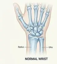
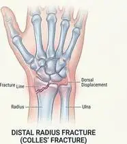
Distal Radius Fracture — Overview
Distal radius fractures (Colles, Smith, and intra-articular Barton fractures) are the most common upper extremity fractures, typically resulting from a fall onto an outstretched hand (FOOSH) with the wrist in dorsiflexion.
A distal radius fracture (wrist fracture) is a break near the wrist end of the larger forearm bone (radius), usually caused by catching yourself with an open hand during a fall.
Primary Causes & Biomechanics
Key Points You Should Know
- •FOOSH mechanism causing dorsal angulation ('dinner-fork' deformity in Colles fracture) and radial shortening
- •Osteopenia/osteoporosis in older adults or high-energy sports/traffic trauma in younger patients
- •Post-immobilization radiocarpal capsular stiffness, flexor/extensor tendon adhesions, and CRPS Type I risk
- •Finger Movement Inside the Cast: Moving your fingers and shoulder while in a cast pumps swelling out and prevents frozen fingers.
- •Wrist & Forearm Turning: Once the cast is removed, gently bending the wrist and turning the palm up/down restores flexibility.
- •Gradual Grip Recovery: Squeezing a soft sponge or therapy ball rebuilds hand strength safely after bone healing.
Assessment Methods
1. Goniometric ROM & Postural / Kinematic Analysis
Wrist active/passive extension (N: 0–70°), flexion (0–80°), ulnar/radial deviation, and forearm supination/pronation (0–85°) measured via goniometry.
2. Special Clinical Provocation & Neurological/Functional Tests
DRUJ ballotment & piano-key test (triangular fibrocartilage complex - TFCC integrity), median nerve carpal tunnel screening, and finger pulp-to-palm distance.
3. Standardized Clinical Outcome Scales & Imaging Criteria
PRWE (Patient-Rated Wrist Evaluation), QuickDASH, Jamar grip & tripod pinch dynamometry, and radiographic radial height/inclination/volar tilt.
1. Range of Motion & Posture Check
Your physiotherapist gently evaluates how comfortably and smoothly your distal radius metaphysis and radiocarpal/distal radioulnar (DRUJ) joints moves and checks your overall posture.
2. Muscle Strength & Functional Movement Check
Your muscle balance, walking or reaching comfort, and specific gentle movement tests are evaluated without triggering sharp pain.
3. Typical Symptoms & Daily Impact Review
Post-cast wrist stiffness, dorsal swelling, and decreased grip strength during daily tasks.
Treatment Options & Phase-Based Protocol
✨ Enriched GIF Exercise & Precaution GuidePhase 1: Acute Symptom Modulation, Tissue Protection & Early Activation
- •Phase 1 Clinical Focus & Tissue Protection: Immobilization Phase (Weeks 0–6): Edema control via elevation, active Six-Pack tendon gliding, full thumb opposition, and active shoulder/elbow ROM every 2 hours while cast/splint is in place to prevent Shoulder-Hand Syndrome (CRPS Type I).
- •Pain, Edema & Neuromuscular Modulation: Targeted physical modalities, relative load management, and early protected mobility of the distal radius metaphysis and radiocarpal/distal radioulnar (DRUJ) joints without provoking mechanical or inflammatory irritation.
- •Proximal & Distal Kinetic Chain Maintenance: Maintain active mobility and circulation in adjacent joints while protecting the primary healing structures in Distal Radius Fracture.
Phase 2: Progressive Mobility, Motor Control & Targeted Strengthening
- •Phase 2 Progressive Mobilization & Motor Control: Early Post-Cast Mobilization (Weeks 6–8): Warm whirlpool/paraffin bath, gentle Maitland Grade I–II radiocarpal/midcarpal glides (convex-concave rule), active/active-assisted wrist flexion-extension, and isolated forearm supination-pronation.
- •Biomechanical Alignment & Co-Contraction: Restore balanced agonist-antagonist force couples, joint centration, and multi-planar neuromuscular control across the distal radius metaphysis and radiocarpal/distal radioulnar (DRUJ) joints.
- •Myofascial Extensibility & Endurance: Address secondary compensatory tightness and build local muscular endurance within safe, symptom-free biomechanical parameters.
Phase 3: Functional Integration, Proprioception & Long-Term Prevention
- •Phase 3 Functional Integration & Long-Term Prevention: Strengthening & Functional Return (Week 8+): Progressive isotonic wrist curls/extensions, Jamar/putty grip strengthening in 20° wrist extension, and closed-kinetic-chain wrist proprioception.
- •Proprioceptive Balance & Dynamic Loading: Progress to task-specific closed-chain, balance, and functional movement patterns tailored to daily life, occupational, or athletic demands.
- •Self-Management & Relapse Prophylaxis: Establish lifelong ergonomic habits, home exercise maintenance, and early warning-sign awareness to prevent recurrence.
Step 1: Calming Pain, Protecting the Area & Gentle Starter Motion
- •Finger Movement Inside the Cast: Moving your fingers and shoulder while in a cast pumps swelling out and prevents frozen fingers.
- •Calm Protection & Gentle Start: During the initial stage of Wrist Fracture, avoid painful overloading and perform the gentle, pain-free starter movements shown below to support circulation and comfort.
- •Respecting Your Body's Signals: Every movement should stay within a comfortable, non-painful zone—never force through sharp or radiating pain.
Step 2: Restoring Flexibility & Building Supporting Muscle Strength
- •Wrist & Forearm Turning: Once the cast is removed, gently bending the wrist and turning the palm up/down restores flexibility.
- •Step-by-Step Flexibility & Support: As acute sensitivity settles, gradually build mobility and strengthen the supporting muscles around the joint at a steady, controlled pace.
- •Posture & Breathing Coordination: Keep your posture upright and breathe smoothly (never hold your breath!) during every repetition.
Step 3: Safe Daily Habits, Stamina & Preventing Recurrence
- •Gradual Grip Recovery: Squeezing a soft sponge or therapy ball rebuilds hand strength safely after bone healing.
- •Confident Return to Daily Life: Integrate your stronger muscles and improved balance into walking, stairs, work, and recreational activities.
- •Preventing Future Flare-Ups: Continue your core protective habits and key maintenance exercises even after symptoms resolve.
Animated (GIF) Therapeutic Exercise Guide
Animated clinical exercise protocol with biomechanical angles, target muscle groups, and dosage parameters (Click image to enlarge)
Step-by-step animated exercise demonstrations you can safely perform at home (Click image to enlarge)
 🔍 Enlarge
1. Six-Pack Differential Flexor Tendon Gliding (FDS & FDP Excursion)
1. Step-by-Step Finger Tendon Gliding (Hook, Straight & Full Fist)
Sequential progression through Straight Hand, Tabletop (intrinsic-plus), Hook Fist (maximum FDS/FDP differential glide), Full Fist, and Straight Fist positions. Prevents peritendinous adhesions within the carpal tunnel and fibro-osseous digital sheaths. Specifically indicated in Distal Radius Fracture rehabilitation to optimize distal radius metaphysis and radiocarpal/distal radioulnar (DRUJ) joints biomechanics and neuromuscular control.
Start with your fingers straight, then bend at the knuckles (tabletop), curl just the fingertips (hook fist), make a gentle full fist, and open wide again—holding each position for 2–3 seconds. Perform smoothly within a comfortable, pain-free range to support your Wrist Fracture recovery.
 🔍 Enlarge
2. Radiocarpal & Midcarpal Active/Eccentric Flexion and Extension
2. Table-Edge Wrist Bending Up and Down
Controlled sagittal plane wrist flexion and extension with the forearm supported on a table edge and digits relaxed. Restores radiocarpal/midcarpal roll-and-glide mechanics and stimulates aligned collagen synthesis in forearm tendons. Specifically indicated in Distal Radius Fracture rehabilitation to optimize distal radius metaphysis and radiocarpal/distal radioulnar (DRUJ) joints biomechanics and neuromuscular control.
Rest your forearm on a table with your wrist hanging comfortably over the edge. Slowly lift your hand upward toward the ceiling, hold for 3 seconds, then lower it gently downward. Perform smoothly within a comfortable, pain-free range to support your Wrist Fracture recovery.
 🔍 Enlarge
3. Proximal & Distal Radioulnar Joint (PRUJ/DRUJ) Supination and Pronation
3. Elbow-at-Side Palm Up and Palm Down Rotation
Isolated forearm rotation with the elbow flexed at 90° and adducted against the trunk to eliminate compensatory glenohumeral rotation. Restores radial head spinning and distal radioulnar translation. Specifically indicated in Distal Radius Fracture rehabilitation to optimize distal radius metaphysis and radiocarpal/distal radioulnar (DRUJ) joints biomechanics and neuromuscular control.
Keep your elbow bent at a right angle and tucked closely against your waist. Slowly turn your palm upward toward the ceiling, hold for 3 seconds, then turn your palm downward toward the floor. Perform smoothly within a comfortable, pain-free range to support your Wrist Fracture recovery.
 🔍 Enlarge
4. Progressive Isotonic Grip & Pinch Strengthening with Extensor Co-Contraction
4. Soft Therapy Ball / Putty Squeeze
Graded isometric squeeze of therapeutic putty or a soft ball with the wrist held in 15°–20° functional extension. Rebuilds extrinsic digital flexor strength alongside synergistic wrist extensor stabilization. Specifically indicated in Distal Radius Fracture rehabilitation to optimize distal radius metaphysis and radiocarpal/distal radioulnar (DRUJ) joints biomechanics and neuromuscular control.
Hold a soft therapy ball or sponge in your palm while keeping your wrist straight. Squeeze gently without triggering sharp pain, hold for 5 seconds, and slowly relax your fingers. Perform smoothly within a comfortable, pain-free range to support your Wrist Fracture recovery.
 🔍 Enlarge
5. Thenar Opposition & Carpometacarpal (CMC) Fine Motor Coordination
5. Thumb-to-Fingertip Touching Sequence
Sequential opposition of the thumb pad to the tips of the index, middle, ring, and little fingers forming an 'O' ring, followed by sliding down the 5th digit toward the distal palmar crease. Restores first web-space mobility and intrinsic precision pinch. Specifically indicated in Distal Radius Fracture rehabilitation to optimize distal radius metaphysis and radiocarpal/distal radioulnar (DRUJ) joints biomechanics and neuromuscular control.
Gently touch the tip of your thumb to each fingertip one by one from your index finger to your little finger, forming a neat 'O' shape each time, then open your hand wide. Perform smoothly within a comfortable, pain-free range to support your Wrist Fracture recovery.
 🔍 Enlarge
6. Biarticular Forearm Flexor & Extensor Myofascial Stretch
6. Straight-Arm Gentle Wrist Flexor & Extensor Stretch
With the elbow in full extension, using the contralateral hand to apply a slow, sustained passive stretch into wrist flexion (for ECRB/extensors) and wrist extension (for common flexors). Reduces resting myotendinous tension. Specifically indicated in Distal Radius Fracture rehabilitation to optimize distal radius metaphysis and radiocarpal/distal radioulnar (DRUJ) joints biomechanics and neuromuscular control.
Extend your arm forward with your elbow straight. Use your other hand to gently bend your wrist downward until you feel a mild stretch in your forearm, hold for 20–30 seconds, then repeat bending upward. Perform smoothly within a comfortable, pain-free range to support your Wrist Fracture recovery.
Movements to Avoid & Strict Precautions
1. Tight Cast Compression & Hanging the Arm Downward
1. Letting your casted arm hang down at your side for hours (causes severe finger swelling!)
2. Forceful Passive Wrist Twisting Before Radiographic Union
2. Forcing or wrenching your stiff wrist painfully right after cast removal
3. Heavy Lifting or Push-Ups on an Unhealed Wrist
3. Lifting heavy pots/weights or leaning full body weight on your hand early on
4. Ignoring Shoulder & Finger Movement During Cast Immobilization
4. Keeping your fingers and shoulder frozen while wearing the wrist cast
Distal Radius Fracture — Video Guide
Click directly on any video to open and watch in a window, or review details via 📋 Summary & Options.
Condition Overview Video (1 Video)
Summary of pathophysiology, biomechanics, and clinical presentation
What the condition is, why it develops, and its basic mechanism

What Is a Wrist Fracture (Distal Radius Fracture)? How Is It Treated?
Distal radius fractures (Colles, Smith, and intra-articular Barton fractures) are the most common upper extremity fractures, typically resulting from a fall onto an outstretched hand (FOOSH) with the wrist in dorsiflexion.
A distal radius fracture (wrist fracture) is a break near the wrist end of the larger forearm bone (radius), usually caused by catching yourself with an open hand during a fall.
Treatment and Exercise Videos
Phase-based physiotherapy protocols, mobilization, and therapeutic exercises
Step-by-step recovery and safe home exercise applications
Wrist Fracture Exercises | Home Physical Therapy and Edema Control Routine
Phase 1: Acute Symptom Modulation, Tissue Protection & Early Activation: Phase 1 Clinical Focus & Tissue Protection: Immobilization Phase (Weeks 0–6): Edema control via elevation, active Six-Pack tendon gliding, full thumb opposition, and active shoulder/elbow ROM every 2 hours while cast/splint is in place to prevent Shoulder-Hand Syndrome (CRPS Type I).
Step 1: Calming Pain, Protecting the Area & Gentle Starter Motion: Finger Movement Inside the Cast: Moving your fingers and shoulder while in a cast pumps swelling out and prevents frozen fingers.

Wrist Fracture Rehabilitation and Range of Motion Exercises
Phase 2: Progressive Mobility, Motor Control & Targeted Strengthening: Phase 2 Progressive Mobilization & Motor Control: Early Post-Cast Mobilization (Weeks 6–8): Warm whirlpool/paraffin bath, gentle Maitland Grade I–II radiocarpal/midcarpal glides (convex-concave rule), active/active-assisted wrist flexion-extension, and isolated forearm supination-pronation.
Step 2: Restoring Flexibility & Building Supporting Muscle Strength: Wrist & Forearm Turning: Once the cast is removed, gently bending the wrist and turning the palm up/down restores flexibility.
Clinical Assessment, Goniometry & Special Test Videos
Displayed only in Healthcare Professional mode (hidden in Patient mode)

Wrist Joint Goniometric Measurement (Flexion, Extension, Radial/Ulnar Deviation)
Goniometric ROM & Postural / Kinematic Analysis: Wrist active/passive extension (N: 0–70°), flexion (0–80°), ulnar/radial deviation, and forearm supination/pronation (0–85°) measured via goniometry.

Wrist Clinical Examination and Functional Assessment
Special Clinical Provocation & Neurological/Functional Tests: DRUJ ballotment & piano-key test (triangular fibrocartilage complex - TFCC integrity), median nerve carpal tunnel screening, and finger pulp-to-palm distance.

Grip Strength Assessment Using a Jamar Hand Dynamometer
Standardized Clinical Outcome Scales & Imaging Criteria: PRWE (Patient-Rated Wrist Evaluation), QuickDASH, Jamar grip & tripod pinch dynamometry, and radiographic radial height/inclination/volar tilt.