Developmental Dysplasia of the Hip (DDH)Congenital Hip Dislocation (Developmental Dysplasia of the Hip)
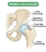
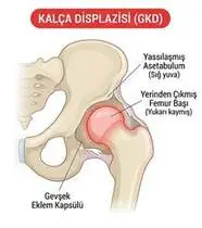
Developmental Dysplasia of the Hip (DDH) — Overview
Developmental Dysplasia of the Hip (DDH) in adolescents and adults encompasses a spectrum from residual acetabular dysplasia (shallow, obliquely oriented acetabular roof with Lateral Center-Edge Angle <20° resulting in deficient anterolateral coverage of the femoral head, excessive labral shear stress, and functional micro-instability) to secondary dysplastic coxarthrosis. In adults, the iliopsoas and dynamic pericapsular muscles (gluteus medius, minimus, maximus, and deep external rotators) must work as active dynamic stabilizers to keep the femoral head centered.
Developmental dysplasia of the hip (DDH / shallow hip socket) is a condition beginning at birth where the roof of the hip socket (acetabulum) is shallower than normal, covering less of the ball of the thighbone. In teens and adults, strengthening the deep hip and buttock muscles acts like a muscular cup that holds the ball centered and protects the joint cartilage.
Primary Causes & Biomechanics
Key Points You Should Know
- •Incomplete remodeling of congenital/infantile hip dysplasia or unrecognized mild acetabular dysplasia presenting in young adulthood (ages 15–35, 80% female)
- •Reduced weight-bearing acetabular contact area concentrating mechanical stress on the hypertrophied anterosuperior labrum and cartilage rim
- •Shortened abductor lever arm (coxa valga and increased femoral anteversion) causing early gluteus medius fatigue and Trendelenburg gait
- •Muscles Acting as a Deeper Socket: Because the bony roof is shallow, your outer hip muscles (gluteus medius) are your hip's best friend—keeping them strong stops the ball from sliding against the rim.
- •Avoid Extreme Hip Stretches: Even if your hips feel very flexible, avoid doing extreme splits or forcing your hip backward, which stretches the joint capsule.
- •Stopping the Side-to-Side Sway (Trendelenburg): Standing balance and side leg lifts train your pelvis to stay level when you walk.
Assessment Methods
1. Goniometric ROM & Postural / Kinematic Analysis
Hip ROM (typically showing excessive internal rotation due to femoral anteversion in young dysplasia, transitioning to internal rotation loss once secondary OA develops).
2. Special Clinical Provocation & Neurological/Functional Tests
Anterior Apprehension / Hyperextension-External Rotation (HEER) Test (anterior micro-instability!), Abduction-Extension-External Rotation Test, FADIR Test (labral overload), and Trendelenburg Sign.
3. Standardized Clinical Outcome Scales & Imaging Criteria
Lateral Center-Edge Angle of Wiberg (LCEA <20° confirms dysplasia), Tönnis roof angle (>10°), Harris Hip Score / iHOT-12, and hip MRI/arthroplasty/periacetabular osteotomy (PAO) planning.
1. Range of Motion & Posture Check
Your physiotherapist gently evaluates how comfortably and smoothly your acetabulum (shallow dysplastic roof, labrum) and proximal femur (femoral head coverage, anteversion, and abductor lever arm) moves and checks your overall posture.
2. Muscle Strength & Functional Movement Check
Your muscle balance, walking or reaching comfort, and specific gentle movement tests are evaluated without triggering sharp pain.
3. Typical Symptoms & Daily Impact Review
Deep aching pain in the groin and outer hip (lateral trochanteric fatigue) after walking or standing, often accompanied by a feeling of the hip catching or 'giving way'.
Treatment Options & Phase-Based Protocol
✨ Enriched GIF Exercise & Precaution GuidePhase 1: Acute Symptom Modulation, Tissue Protection & Early Activation
- •Phase 1 Clinical Focus & Tissue Protection: Dynamic Joint Centering & Isometric Activation Phase (Weeks 1–3): Avoid end-range hip hyperextension and extreme stretching; perform isometric gluteal & quadriceps sets, unloaded supine heel slides, and posterior pelvic tilts.
- •Pain, Edema & Neuromuscular Modulation: Targeted physical modalities, relative load management, and early protected mobility of the acetabulum (shallow dysplastic roof, labrum) and proximal femur (femoral head coverage, anteversion, and abductor lever arm) without provoking mechanical or inflammatory irritation.
- •Proximal & Distal Kinetic Chain Maintenance: Maintain active mobility and circulation in adjacent joints while protecting the primary healing structures in Developmental Dysplasia of the Hip.
Phase 2: Progressive Mobility, Motor Control & Targeted Strengthening
- •Phase 2 Progressive Mobilization & Motor Control: Gluteus Medius & Deep Stabilizer Hypertrophy Phase (Weeks 3–8): Side-lying hip abduction, standing supported hip abduction, supine pelvic bridging, and quadruped Bird-Dog to build the muscular cuff that prevents anterosuperior femoral head migration.
- •Biomechanical Alignment & Co-Contraction: Restore balanced agonist-antagonist force couples, joint centration, and multi-planar neuromuscular control across the acetabulum (shallow dysplastic roof, labrum) and proximal femur (femoral head coverage, anteversion, and abductor lever arm).
- •Myofascial Extensibility & Endurance: Address secondary compensatory tightness and build local muscular endurance within safe, symptom-free biomechanical parameters.
Phase 3: Functional Integration, Proprioception & Long-Term Prevention
- •Phase 3 Functional Integration & Long-Term Prevention: Functional Gait & Post-Osteotomy/THA Phase (Week 8+): Controlled chair sit-to-stand, single-leg pelvic drop control (eliminating Trendelenburg limp!), low-impact cycling/swimming, and post-PAO/arthroplasty weight-bearing progression.
- •Proprioceptive Balance & Dynamic Loading: Progress to task-specific closed-chain, balance, and functional movement patterns tailored to daily life, occupational, or athletic demands.
- •Self-Management & Relapse Prophylaxis: Establish lifelong ergonomic habits, home exercise maintenance, and early warning-sign awareness to prevent recurrence.
Step 1: Calming Pain, Protecting the Area & Gentle Starter Motion
- •Muscles Acting as a Deeper Socket: Because the bony roof is shallow, your outer hip muscles (gluteus medius) are your hip's best friend—keeping them strong stops the ball from sliding against the rim.
- •Calm Protection & Gentle Start: During the initial stage of Congenital Hip Dislocation, avoid painful overloading and perform the gentle, pain-free starter movements shown below to support circulation and comfort.
- •Respecting Your Body's Signals: Every movement should stay within a comfortable, non-painful zone—never force through sharp or radiating pain.
Step 2: Restoring Flexibility & Building Supporting Muscle Strength
- •Avoid Extreme Hip Stretches: Even if your hips feel very flexible, avoid doing extreme splits or forcing your hip backward, which stretches the joint capsule.
- •Step-by-Step Flexibility & Support: As acute sensitivity settles, gradually build mobility and strengthen the supporting muscles around the joint at a steady, controlled pace.
- •Posture & Breathing Coordination: Keep your posture upright and breathe smoothly (never hold your breath!) during every repetition.
Step 3: Safe Daily Habits, Stamina & Preventing Recurrence
- •Stopping the Side-to-Side Sway (Trendelenburg): Standing balance and side leg lifts train your pelvis to stay level when you walk.
- •Confident Return to Daily Life: Integrate your stronger muscles and improved balance into walking, stairs, work, and recreational activities.
- •Preventing Future Flare-Ups: Continue your core protective habits and key maintenance exercises even after symptoms resolve.
Animated (GIF) Therapeutic Exercise Guide
Animated clinical exercise protocol with biomechanical angles, target muscle groups, and dosage parameters (Click image to enlarge)
Step-by-step animated exercise demonstrations you can safely perform at home (Click image to enlarge)
 🔍 Enlarge
1. Supine Active-Assisted Heel Slide & Synovial Diffusion Mobilization
1. Supine Heel Slide (Gentle Knee & Hip Bending)
Controlled flexion and extension of the lower extremity by sliding the heel along the support surface without axial weight-bearing. Stimulates synovial fluid diffusion across avascular cartilage and prevents capsular adhesions. Specifically indicated in Developmental Dysplasia of the Hip rehabilitation to optimize acetabulum (shallow dysplastic roof, labrum) and proximal femur (femoral head coverage, anteversion, and abductor lever arm) biomechanics and neuromuscular control.
While lying on your back, slowly slide your heel along the bed sheet toward your hip within a comfortable, pain-free angle, hold for 3 seconds, and gently slide it back out straight. Perform smoothly within a comfortable, pain-free range to support your Congenital Hip Dislocation recovery.
 🔍 Enlarge
2. Side-Lying Hip Abduction (Gluteus Medius & Frontal Plane Control)
2. Side-Lying Leg Lift (Outer Hip Strengthening)
Isolated strengthening of the gluteus medius in slight hip extension and neutral rotation. Prevents contralateral pelvic drop (Trendelenburg) and reduces dynamic knee valgus and medial compartment adduction moments. Specifically indicated in Developmental Dysplasia of the Hip rehabilitation to optimize acetabulum (shallow dysplastic roof, labrum) and proximal femur (femoral head coverage, anteversion, and abductor lever arm) biomechanics and neuromuscular control.
Lie on your side with your bottom leg slightly bent for balance. Keep your top leg straight and slightly back, lift it up toward the ceiling without rolling your pelvis backward, hold for 3 seconds, and lower slowly. Perform smoothly within a comfortable, pain-free range to support your Congenital Hip Dislocation recovery.
 🔍 Enlarge
3. Supported Standing Hip Abduction & Closed-Chain Stance Stabilization
3. Counter-Supported Standing Side Leg Lift
Standing upright with light hand support, abducting the lower limb in the frontal plane while the stance-leg gluteus medius contracts isometrically to keep the pelvis level. Builds functional gait stability. Specifically indicated in Developmental Dysplasia of the Hip rehabilitation to optimize acetabulum (shallow dysplastic roof, labrum) and proximal femur (femoral head coverage, anteversion, and abductor lever arm) biomechanics and neuromuscular control.
Hold lightly onto a sturdy counter or chair back and stand tall. Without leaning your trunk sideways, lift your leg out to the side, pause for 2–3 seconds, and slowly return. Perform smoothly within a comfortable, pain-free range to support your Congenital Hip Dislocation recovery.
 🔍 Enlarge
4. Functional Closed-Kinetic-Chain Chair Transfer (Sit-to-Stand Biomechanics)
4. Controlled Chair Sit-to-Stand Transfer
Controlled transition from sitting to standing and eccentric lowering back to the chair with symmetrical weight distribution, anterior trunk inclination ('nose over toes'), and neutral frontal knee alignment. Specifically indicated in Developmental Dysplasia of the Hip rehabilitation to optimize acetabulum (shallow dysplastic roof, labrum) and proximal femur (femoral head coverage, anteversion, and abductor lever arm) biomechanics and neuromuscular control.
Sit near the front of a sturdy chair with your feet hip-width apart. Lean slightly forward and push evenly through both heels to stand up tall, then slowly lower yourself back down over 3 seconds. Perform smoothly within a comfortable, pain-free range to support your Congenital Hip Dislocation recovery.
 🔍 Enlarge
5. Supine Lumbopelvic Bridging (Gluteus Maximus & Posterior Chain Co-Contraction)
5. Supine Hip Bridge (Controlled Pelvic Lift)
From hook-lying with transversus abdominis pre-activation, elevating the pelvis until hips, pelvis, and shoulders form a neutral diagonal line. Strengthens the gluteus maximus, hamstrings, and erector spinae synergy without lumbar hyperlordosis. Specifically indicated in Developmental Dysplasia of the Hip rehabilitation to optimize acetabulum (shallow dysplastic roof, labrum) and proximal femur (femoral head coverage, anteversion, and abductor lever arm) biomechanics and neuromuscular control.
Lie on your back with your knees bent and feet flat hip-width apart. Gently tighten your tummy and glutes, lift your hips until your body forms a straight line from shoulders to knees, hold for 5 seconds, and lower slowly. Perform smoothly within a comfortable, pain-free range to support your Congenital Hip Dislocation recovery.
 🔍 Enlarge
6. Quadruped Contralateral Arm-Leg Reach ('Bird-Dog' Multifidus & Deep Core)
6. All-Fours Opposite Arm & Leg Reach (Bird-Dog)
In quadruped with a neutral lumbopelvic spine and abdominal drawing-in (transversus abdominis), extending opposite upper and lower extremities horizontally without pelvic rotation or lumbar hyperextension. Minimizes spinal compressive load while maximizing multifidus recruitment. Specifically indicated in Developmental Dysplasia of the Hip rehabilitation to optimize acetabulum (shallow dysplastic roof, labrum) and proximal femur (femoral head coverage, anteversion, and abductor lever arm) biomechanics and neuromuscular control.
On your hands and knees, keep your back flat like a tabletop and gently tighten your tummy. Slowly reach one arm forward and the opposite leg straight back withoutarching your lower back, hold for 5 seconds, and switch sides. Perform smoothly within a comfortable, pain-free range to support your Congenital Hip Dislocation recovery.
Movements to Avoid & Strict Precautions
1. Extreme Passive Hip Stretching (Splits, Forced Frog-Stretches, Extreme Yoga Backbends)
1. Doing splits, extreme hip-opening yoga poses, or forcing your leg far backward (causes micro-instability!)
2. Standing With All Weight Hanging on One Hip ('Hanging on the Ligaments')
2. Standing with your hip popped out to one side hanging on the joint capsule
3. High-Impact Long-Distance Running on Asphalt or Heavy Jumping
3. Long-distance running on concrete or high-impact jumping
4. Crossing the Legs or Bending Past 90° After Total Hip Arthroplasty
4. Crossing your legs or sitting on low seats after hip replacement surgery
Developmental Dysplasia of the Hip (DDH) — Video Guide
Click directly on any video to open and watch in a window, or review details via 📋 Summary & Options.
Condition Overview Video (1 Video)
Summary of pathophysiology, biomechanics, and clinical presentation
What the condition is, why it develops, and its basic mechanism
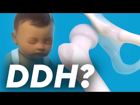
What Is Developmental Dysplasia of the Hip (DDH)? Diagnosis and Treatment
Developmental Dysplasia of the Hip (DDH) in adolescents and adults encompasses a spectrum from residual acetabular dysplasia (shallow, obliquely oriented acetabular roof with Lateral Center-Edge Angle <20° resulting in deficient anterolateral coverage of the femoral head, excessive labral shear stress, and functional micro-instability) to secondary dysplastic coxarthrosis. In adults, the iliopsoas and dynamic pericapsular muscles (gluteus medius, minimus, maximus, and deep external rotators) must work as active dynamic stabilizers to keep the femoral head centered.
Developmental dysplasia of the hip (DDH / shallow hip socket) is a condition beginning at birth where the roof of the hip socket (acetabulum) is shallower than normal, covering less of the ball of the thighbone. In teens and adults, strengthening the deep hip and buttock muscles acts like a muscular cup that holds the ball centered and protects the joint cartilage.
Treatment and Exercise Videos
Phase-based physiotherapy protocols, mobilization, and therapeutic exercises
Step-by-step recovery and safe home exercise applications

Early Diagnosis, Bracing, and Rehabilitation Guide in Developmental Hip Dysplasia
Phase 1: Acute Symptom Modulation, Tissue Protection & Early Activation: Phase 1 Clinical Focus & Tissue Protection: Dynamic Joint Centering & Isometric Activation Phase (Weeks 1–3): Avoid end-range hip hyperextension and extreme stretching; perform isometric gluteal & quadriceps sets, unloaded supine heel slides, and posterior pelvic tilts.
Step 1: Calming Pain, Protecting the Area & Gentle Starter Motion: Muscles Acting as a Deeper Socket: Because the bony roof is shallow, your outer hip muscles (gluteus medius) are your hip's best friend—keeping them strong stops the ball from sliding against the rim.

Clinical Tests and Therapeutic Exercises in Hip and Groin Pain
Phase 2: Progressive Mobility, Motor Control & Targeted Strengthening: Phase 2 Progressive Mobilization & Motor Control: Gluteus Medius & Deep Stabilizer Hypertrophy Phase (Weeks 3–8): Side-lying hip abduction, standing supported hip abduction, supine pelvic bridging, and quadruped Bird-Dog to build the muscular cuff that prevents anterosuperior femoral head migration.
Step 2: Restoring Flexibility & Building Supporting Muscle Strength: Avoid Extreme Hip Stretches: Even if your hips feel very flexible, avoid doing extreme splits or forcing your hip backward, which stretches the joint capsule.
Clinical Assessment, Goniometry & Special Test Videos
Displayed only in Healthcare Professional mode (hidden in Patient mode)

Barlow and Ortolani Tests — Developmental Dysplasia of the Hip Examination
Goniometric ROM & Postural / Kinematic Analysis: Hip ROM (typically showing excessive internal rotation due to femoral anteversion in young dysplasia, transitioning to internal rotation loss once secondary OA develops).

Developmental Hip Dysplasia (DDH) Clinical Hip Abduction and Instability Exam
Special Clinical Provocation & Neurological/Functional Tests: Anterior Apprehension / Hyperextension-External Rotation (HEER) Test (anterior micro-instability!), Abduction-Extension-External Rotation Test, FADIR Test (labral overload), and Trendelenburg Sign.

Hip Joint Goniometric Measurement (Flexion, Extension, Abduction, Rotations)
Standardized Clinical Outcome Scales & Imaging Criteria: Lateral Center-Edge Angle of Wiberg (LCEA <20° confirms dysplasia), Tönnis roof angle (>10°), Harris Hip Score / iHOT-12, and hip MRI/arthroplasty/periacetabular osteotomy (PAO) planning.