Calcaneal Spur (Heel Spur)Heel Spur (Calcaneal Spur)
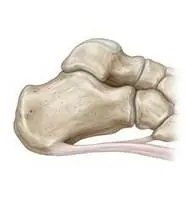
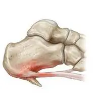
Calcaneal Spur (Heel Spur) — Overview
A calcaneal spur (heel spur) is a reactive shelf-like traction osteophyte (bony outgrowth ≥2 mm) forming at the medial process of the calcaneal tuberosity (typically deep in the origin of the *M. flexor digitorum brevis* and plantar aponeurosis) in response to chronic tensile traction and micro-trauma. Crucially, the pain stems from the surrounding inflamed plantar enthesis, periosteum, Baxter's nerve irritation, and heel fat-pad micro-trauma rather than the bony spur tip itself pointing vertically downward.
A calcaneal spur (heel spur) is a small calcium bone projection that forms on the underside of your heel bone after months of chronic pulling by the foot-arch band and muscles. The good news is that you do not need surgery to shave the bone off—once we stretch the tight calf/arch and cushion the heel, the tissue inflammation heals completely even though the little spur stays on X-ray!
Primary Causes & Biomechanics
Key Points You Should Know
- •Chronic unaddressed plantar fasciopathy and repetitive traction of the flexor digitorum brevis / plantar aponeurosis on the calcaneal periosteum
- •Tight gastrocnemius-soleus-Achilles complex and age-related atrophy of the shock-absorbing heel fat pad
- •Prolonged standing on hard surfaces, elevated BMI, and pes planus/cavus biomechanical overload
- •Why the Spur Doesn't Need to Be Removed: Millions of pain-free people have a heel spur on X-ray! The pain comes from the swollen tissue pulled tight around it—relaxing that tension cures the pain.
- •Silicone Heel Cup with a Soft Center: Using a silicone heel pad with a softer center spot takes direct body-weight pressure off the sensitive heel point.
- •Calf Stretching Is Half the Cure: A tight calf muscle pulls on the back of the heel bone, which pulls directly on the bottom heel spur with every step.
Assessment Methods
1. Goniometric ROM & Postural / Kinematic Analysis
Ankle dorsiflexion goniometry (typically <5–10° with knee extended) and 1st MTP extension.
2. Special Clinical Provocation & Neurological/Functional Tests
Central-medial plantar heel palpation, Windlass test, Calcaneal Squeeze test (differential for stress fracture), and Heel Fat Pad thickness assessment.
3. Standardized Clinical Outcome Scales & Imaging Criteria
Lateral weight-bearing foot radiograph (demonstrating the inferior calcaneal osteophyte), FFI, and FAAM.
1. Range of Motion & Posture Check
Your physiotherapist gently evaluates how comfortably and smoothly your inferior calcaneal tuberosity (traction osteophyte at flexor digitorum brevis / plantar fascia origin) and heel fat pad moves and checks your overall posture.
2. Muscle Strength & Functional Movement Check
Your muscle balance, walking or reaching comfort, and specific gentle movement tests are evaluated without triggering sharp pain.
3. Typical Symptoms & Daily Impact Review
Sharp, bruised, or 'stepping-on-a-stone/nail' pain under the center-inner heel during the first steps in the morning and after sitting.
Treatment Options & Phase-Based Protocol
✨ Enriched GIF Exercise & Precaution GuidePhase 1: Acute Symptom Modulation, Tissue Protection & Early Activation
- •Phase 1 Clinical Focus & Tissue Protection: Heel Offloading, Physical Modalities & Morning Stretch Phase (Weeks 1–3): Silicone heel cup with central soft-spot cutout (offloads the spur zone by 40%), ESWT (Extracorporeal Shockwave Therapy — radial/focused) or therapeutic ultrasound, ice-bottle rolling, and pre-step morning towel calf & plantar fascia stretches.
- •Pain, Edema & Neuromuscular Modulation: Targeted physical modalities, relative load management, and early protected mobility of the inferior calcaneal tuberosity (traction osteophyte at flexor digitorum brevis / plantar fascia origin) and heel fat pad without provoking mechanical or inflammatory irritation.
- •Proximal & Distal Kinetic Chain Maintenance: Maintain active mobility and circulation in adjacent joints while protecting the primary healing structures in Calcaneal Spur.
Phase 2: Progressive Mobility, Motor Control & Targeted Strengthening
- •Phase 2 Progressive Mobilization & Motor Control: Intrinsic Arch Support & Calf Lengthening Phase (Weeks 3–6): Short-Foot arch doming & towel curls (unloading the calcaneal enthesis by activating intrinsic muscles), ankle pumps, and daily gastro-soleus stretching.
- •Biomechanical Alignment & Co-Contraction: Restore balanced agonist-antagonist force couples, joint centration, and multi-planar neuromuscular control across the inferior calcaneal tuberosity (traction osteophyte at flexor digitorum brevis / plantar fascia origin) and heel fat pad.
- •Myofascial Extensibility & Endurance: Address secondary compensatory tightness and build local muscular endurance within safe, symptom-free biomechanical parameters.
Phase 3: Functional Integration, Proprioception & Long-Term Prevention
- •Phase 3 Functional Integration & Long-Term Prevention: Progressive Tendon-Fascia Remodeling Phase (Week 6–12): Controlled bilateral heel raises (calf raises), balance/weight-shift training, and transition to rocker-bottom or cushioned footwear with custom arch orthotics.
- •Proprioceptive Balance & Dynamic Loading: Progress to task-specific closed-chain, balance, and functional movement patterns tailored to daily life, occupational, or athletic demands.
- •Self-Management & Relapse Prophylaxis: Establish lifelong ergonomic habits, home exercise maintenance, and early warning-sign awareness to prevent recurrence.
Step 1: Calming Pain, Protecting the Area & Gentle Starter Motion
- •Why the Spur Doesn't Need to Be Removed: Millions of pain-free people have a heel spur on X-ray! The pain comes from the swollen tissue pulled tight around it—relaxing that tension cures the pain.
- •Calm Protection & Gentle Start: During the initial stage of Heel Spur, avoid painful overloading and perform the gentle, pain-free starter movements shown below to support circulation and comfort.
- •Respecting Your Body's Signals: Every movement should stay within a comfortable, non-painful zone—never force through sharp or radiating pain.
Step 2: Restoring Flexibility & Building Supporting Muscle Strength
- •Silicone Heel Cup with a Soft Center: Using a silicone heel pad with a softer center spot takes direct body-weight pressure off the sensitive heel point.
- •Step-by-Step Flexibility & Support: As acute sensitivity settles, gradually build mobility and strengthen the supporting muscles around the joint at a steady, controlled pace.
- •Posture & Breathing Coordination: Keep your posture upright and breathe smoothly (never hold your breath!) during every repetition.
Step 3: Safe Daily Habits, Stamina & Preventing Recurrence
- •Calf Stretching Is Half the Cure: A tight calf muscle pulls on the back of the heel bone, which pulls directly on the bottom heel spur with every step.
- •Confident Return to Daily Life: Integrate your stronger muscles and improved balance into walking, stairs, work, and recreational activities.
- •Preventing Future Flare-Ups: Continue your core protective habits and key maintenance exercises even after symptoms resolve.
Animated (GIF) Therapeutic Exercise Guide
Animated clinical exercise protocol with biomechanical angles, target muscle groups, and dosage parameters (Click image to enlarge)
Step-by-step animated exercise demonstrations you can safely perform at home (Click image to enlarge)
 🔍 Enlarge
1. Long-Sitting Towel-Assisted Gastrocnemius-Soleus & Plantar Fascia Stretch
1. Seated Towel Calf & Foot Arch Stretch
Long-sitting with a towel looped around the metatarsal heads, applying a slow, sustained passive pull into ankle dorsiflexion. Restores gastrocnemius-soleus-Achilles length and reduces plantar fascial and equine tension. Specifically indicated in Calcaneal Spur rehabilitation to optimize inferior calcaneal tuberosity (traction osteophyte at flexor digitorum brevis / plantar fascia origin) and heel fat pad biomechanics and neuromuscular control.
Sit with your leg extended and loop a towel around the ball of your foot. Gently pull both ends of the towel toward you until you feel a comfortable stretch in your calf and sole, holding for 20–30 seconds. Perform smoothly within a comfortable, pain-free range to support your Heel Spur recovery.
 🔍 Enlarge
2. Intrinsic Foot Core ('Short Foot') & Medial Longitudinal Arch Activation
2. Seated Arch Doming (Short Foot) & Towel Scrunch
Drawing the first metatarsal head toward the calcaneus to elevate the medial longitudinal arch without curling the toes (or performing controlled towel curls). Strengthens the intrinsic plantar muscles supporting the subtalar and midtarsal joints. Specifically indicated in Calcaneal Spur rehabilitation to optimize inferior calcaneal tuberosity (traction osteophyte at flexor digitorum brevis / plantar fascia origin) and heel fat pad biomechanics and neuromuscular control.
While seated with your foot flat on the floor (or on a small towel), gently draw the ball of your foot toward your heel to lift your inner arch without curling your toes, hold for 5 seconds, and relax. Perform smoothly within a comfortable, pain-free range to support your Heel Spur recovery.
 🔍 Enlarge
3. Rhythmic Active Ankle Dorsiflexion & Plantarflexion Pumping (Venous/Edema Pump)
3. Ankle Toe-Pulls and Pointing (Circulation & Swelling Pump)
Rhythmic alternating contraction of the tibialis anterior and gastrocnemius-soleus complex. Activates the deep venous calf pump to prevent deep vein thrombosis (DVT), clears distal edema, and maintains talocrural mobility. Specifically indicated in Calcaneal Spur rehabilitation to optimize inferior calcaneal tuberosity (traction osteophyte at flexor digitorum brevis / plantar fascia origin) and heel fat pad biomechanics and neuromuscular control.
While lying down or sitting with your legs supported, pull your toes and feet firmly upward toward your shins, hold for 3 seconds, then point them downward like pressing a gas pedal. Perform smoothly within a comfortable, pain-free range to support your Heel Spur recovery.
 🔍 Enlarge
4. Closed-Chain Bilateral/Eccentric Heel Raise (Triceps Surae & Achilles Loading)
4. Supported Standing Heel Raise & Slow Lowering
Controlled concentric heel elevation followed by slow (3–4 second) eccentric lowering into neutral dorsiflexion. Stimulates Type I collagen alignment in the Achilles/plantar aponeurosis and builds ankle plantarflexor propulsion. Specifically indicated in Calcaneal Spur rehabilitation to optimize inferior calcaneal tuberosity (traction osteophyte at flexor digitorum brevis / plantar fascia origin) and heel fat pad biomechanics and neuromuscular control.
Hold lightly onto a wall or chair back. Rise up onto the balls of both feet, pause at the top, and slowly lower your heels back down to the floor over 3–4 seconds. Perform smoothly within a comfortable, pain-free range to support your Heel Spur recovery.
 🔍 Enlarge
5. Supported Standing Hip Abduction & Closed-Chain Stance Stabilization
5. Counter-Supported Standing Side Leg Lift
Standing upright with light hand support, abducting the lower limb in the frontal plane while the stance-leg gluteus medius contracts isometrically to keep the pelvis level. Builds functional gait stability. Specifically indicated in Calcaneal Spur rehabilitation to optimize inferior calcaneal tuberosity (traction osteophyte at flexor digitorum brevis / plantar fascia origin) and heel fat pad biomechanics and neuromuscular control.
Hold lightly onto a sturdy counter or chair back and stand tall. Without leaning your trunk sideways, lift your leg out to the side, pause for 2–3 seconds, and slowly return. Perform smoothly within a comfortable, pain-free range to support your Heel Spur recovery.
 🔍 Enlarge
6. Single-Leg / Semi-Tandem Proprioceptive & Weight-Shift Balance Training
6. Supported Weight-Shifting & Tandem Balance Practice
Controlled mediolateral/anteroposterior weight shifting and tandem or single-leg stance near a stable support. Enhances mechanoreceptor feedback, ankle/hip postural reflexes, and dynamic gait stability. Specifically indicated in Calcaneal Spur rehabilitation to optimize inferior calcaneal tuberosity (traction osteophyte at flexor digitorum brevis / plantar fascia origin) and heel fat pad biomechanics and neuromuscular control.
Stand near a counter or sturdy chair for safety. Place one foot slightly in front of the other (or shift your weight smoothly from side to side) and hold your balance upright for 20–30 seconds. Perform smoothly within a comfortable, pain-free range to support your Heel Spur recovery.
Movements to Avoid & Strict Precautions
1. Walking Barefoot on Tile/Hardwood Floors or Wearing Hard Flat Shoes
1. Walking barefoot at home or wearing flat, unpadded shoes
2. Repeated Cortisone Injections Into the Heel Fat Pad (Dissolves Your Natural Cushion!)
2. Getting repeated cortisone shots into the bottom of the heel (thins out your natural heel fat pad!)
3. Stepping Out of Bed in the Morning Without Stretching the Calf & Foot Arch First
3. Stepping onto the floor in the morning before stretching your calf and foot in bed
4. Pounding the Heel During High-Impact Running or Jumping on Concrete
4. Running or jumping on concrete with hard heels
Calcaneal Spur (Heel Spur) — Video Guide
Click directly on any video to open and watch in a window, or review details via 📋 Summary & Options.
Condition Overview Video (1 Video)
Summary of pathophysiology, biomechanics, and clinical presentation
What the condition is, why it develops, and its basic mechanism

Calcaneal Spur (Heel Spur) Biomechanics and Relieving Exercise Solutions
A calcaneal spur (heel spur) is a reactive shelf-like traction osteophyte (bony outgrowth ≥2 mm) forming at the medial process of the calcaneal tuberosity (typically deep in the origin of the *M. flexor digitorum brevis* and plantar aponeurosis) in response to chronic tensile traction and micro-trauma. Crucially, the pain stems from the surrounding inflamed plantar enthesis, periosteum, Baxter's nerve irritation, and heel fat-pad micro-trauma rather than the bony spur tip itself pointing vertically downward.
A calcaneal spur (heel spur) is a small calcium bone projection that forms on the underside of your heel bone after months of chronic pulling by the foot-arch band and muscles. The good news is that you do not need surgery to shave the bone off—once we stretch the tight calf/arch and cushion the heel, the tissue inflammation heals completely even though the little spur stays on X-ray!
Treatment and Exercise Videos
Phase-based physiotherapy protocols, mobilization, and therapeutic exercises
Step-by-step recovery and safe home exercise applications

Plantar Fasciitis and Heel Spur Treatment: Pain-Relieving Stretch & Strength Routine
Phase 1: Acute Symptom Modulation, Tissue Protection & Early Activation: Phase 1 Clinical Focus & Tissue Protection: Heel Offloading, Physical Modalities & Morning Stretch Phase (Weeks 1–3): Silicone heel cup with central soft-spot cutout (offloads the spur zone by 40%), ESWT (Extracorporeal Shockwave Therapy — radial/focused) or therapeutic ultrasound, ice-bottle rolling, and pre-step morning towel calf & plantar fascia stretches.
Step 1: Calming Pain, Protecting the Area & Gentle Starter Motion: Why the Spur Doesn't Need to Be Removed: Millions of pain-free people have a heel spur on X-ray! The pain comes from the swollen tissue pulled tight around it—relaxing that tension cures the pain.

Home Therapeutic Exercises for Plantar Fasciitis and Heel Pain
Phase 2: Progressive Mobility, Motor Control & Targeted Strengthening: Phase 2 Progressive Mobilization & Motor Control: Intrinsic Arch Support & Calf Lengthening Phase (Weeks 3–6): Short-Foot arch doming & towel curls (unloading the calcaneal enthesis by activating intrinsic muscles), ankle pumps, and daily gastro-soleus stretching.
Step 2: Restoring Flexibility & Building Supporting Muscle Strength: Silicone Heel Cup with a Soft Center: Using a silicone heel pad with a softer center spot takes direct body-weight pressure off the sensitive heel point.
Clinical Assessment, Goniometry & Special Test Videos
Displayed only in Healthcare Professional mode (hidden in Patient mode)

Ankle and Foot Goniometric Measurement (Dorsiflexion, Plantarflexion, Inversion/Eversion)
Goniometric ROM & Postural / Kinematic Analysis: Ankle dorsiflexion goniometry (typically <5–10° with knee extended) and 1st MTP extension.