Ankylosing Spondylitis (Axial Spondyloarthritis)Inflammatory Spinal Rheumatism (Ankylosing Spondylitis)
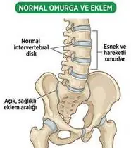
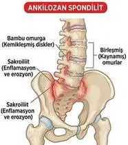
Ankylosing Spondylitis (Axial Spondyloarthritis) — Overview
Ankylosing Spondylitis (radiographic Axial Spondyloarthritis — axSpA) is a chronic HLA-B27–associated inflammatory rheumatic disease targeting the entheses (sites where ligaments, tendons, and joint capsules attach to bone) of the sacroiliac joints and axial skeleton. Chronic enthesitis undergoes ossification into bridging syndesmophytes ('bamboo spine'), leading to loss of lumbar lordosis, thoracic hyperkyphosis, costovertebral ankylosis (restrictive chest wall restriction), and hip flexion contractures. Crucially, daily spinal extension and deep costal expansion exercises are as fundamental as biologic/NSAID therapy!
Ankylosing spondylitis (inflammatory spinal rheumatism) is an inflammatory condition starting in the pelvic joints (sacroiliac joints) and spine in young adults. Unlike mechanical back pain (which feels better with rest), ankylosing spondylitis causes stiffness in the second half of the night and early morning (>45 minutes) that dramatically improves as soon as you exercise and take a warm shower!
Primary Causes & Biomechanics
Key Points You Should Know
- •HLA-B27 genetic association (present in ~90% of patients) and immune-mediated enthesitis starting bilaterally at the sacroiliac joints in young adults (<40 years)
- •Progressive ascending ossification of the annulus fibrosus (syndesmophytes), apophyseal joints, and costovertebral/costotransverse joints
- •Loss of lumbar lordosis + thoracic kyphosis + cervical forward flexion creating the classic 'skier's posture' and restricting forward visual gaze and vital capacity
- •Movement Is Your Medicine: In ankylosing spondylitis, resting makes the spine stiffer, while daily spinal extension and chest-opening exercises keep the vertebrae mobile and upright.
- •The 20-Minute Tummy-Lying Habit: Lying flat on your stomach on a firm mattress for 20 minutes every day (and sleeping on a firm mattress with a very thin pillow) prevents stooped posture.
- •Deep Ribcage Breathing: Because the joints where your ribs meet your spine can stiffen, deep chest-expanding breaths with arm raises protect your lung capacity.
Assessment Methods
1. Goniometric ROM & Postural / Kinematic Analysis
BASMI (Bath Ankylosing Spondylitis Metrology Index)—1) Modified Schober Test (lumbar flexion <4–5 cm expansion indicates restriction), 2) Chest Expansion Difference at the 4th intercostal space (Normal: ≥5 cm; <2.5 cm indicates costovertebral ankylosis!), 3) Tragus-to-Wall / Occiput-to-Wall Distance (Normal: 0 cm), 4) Cervical Rotation, and 5) Intermalleolar Distance (hip abduction).
2. Special Clinical Provocation & Neurological/Functional Tests
Sacroiliac provocation cluster (FABER, Gaenslen's, sacral thrust) and Thomas test (hip flexion contracture).
3. Standardized Clinical Outcome Scales & Imaging Criteria
BASDAI (Disease Activity), BASFI (Functional Index), BASMI, and SIJ/spine MRI (active bone marrow edema sacroiliitis) & radiographs (New York criteria).
1. Range of Motion & Posture Check
Your physiotherapist gently evaluates how comfortably and smoothly your sacroiliac joints, axial spine (intervertebral discs, facet/costovertebral/costotransverse joints — entheses), and hip/shoulder girdle moves and checks your overall posture.
2. Muscle Strength & Functional Movement Check
Your muscle balance, walking or reaching comfort, and specific gentle movement tests are evaluated without triggering sharp pain.
3. Typical Symptoms & Daily Impact Review
Inflammatory Back & Alternating Buttock Pain beginning before age 40: insidious onset (>3 months), morning stiffness lasting >45–60 minutes, awakening in the second half of the night with back stiffness, and marked improvement with exercise/warm shower but no relief with rest.
Treatment Options & Phase-Based Protocol
✨ Enriched GIF Exercise & Precaution GuidePhase 1: Acute Symptom Modulation, Tissue Protection & Early Activation
- •Phase 1 Clinical Focus & Tissue Protection: Daily Postural Extension & Prone Positioning Phase (Lifelong Core Habit): Mandatory 20-minute daily prone lying (flat on the stomach before sleep or in the morning) to prevent hip and thoracic flexion contractures, plus wall-referenced axial spinal extension (pressing heels, hips, shoulders, and occiput to the wall).
- •Pain, Edema & Neuromuscular Modulation: Targeted physical modalities, relative load management, and early protected mobility of the sacroiliac joints, axial spine (intervertebral discs, facet/costovertebral/costotransverse joints — entheses), and hip/shoulder girdle without provoking mechanical or inflammatory irritation.
- •Proximal & Distal Kinetic Chain Maintenance: Maintain active mobility and circulation in adjacent joints while protecting the primary healing structures in Ankylosing Spondylitis.
Phase 2: Progressive Mobility, Motor Control & Targeted Strengthening
- •Phase 2 Progressive Mobilization & Motor Control: Costovertebral Thoracic Expansion Phase: Deep lateral/basal costal breathing with bilateral arm elevation (Thoracic Expansion Exercises) and scapular retraction to preserve chest expansion (>5 cm) and pulmonary vital capacity.
- •Biomechanical Alignment & Co-Contraction: Restore balanced agonist-antagonist force couples, joint centration, and multi-planar neuromuscular control across the sacroiliac joints, axial spine (intervertebral discs, facet/costovertebral/costotransverse joints — entheses), and hip/shoulder girdle.
- •Myofascial Extensibility & Endurance: Address secondary compensatory tightness and build local muscular endurance within safe, symptom-free biomechanical parameters.
Phase 3: Functional Integration, Proprioception & Long-Term Prevention
- •Phase 3 Functional Integration & Long-Term Prevention: Spinal Mobility, Hip Extensor & Aquatic Phase: Quadruped Bird-Dog & cat-camel segmental mobilization, prone active back/hip extension, supine pelvic bridging, and swimming (especially backstroke/freestyle) & hydrotherapy.
- •Proprioceptive Balance & Dynamic Loading: Progress to task-specific closed-chain, balance, and functional movement patterns tailored to daily life, occupational, or athletic demands.
- •Self-Management & Relapse Prophylaxis: Establish lifelong ergonomic habits, home exercise maintenance, and early warning-sign awareness to prevent recurrence.
Step 1: Calming Pain, Protecting the Area & Gentle Starter Motion
- •Movement Is Your Medicine: In ankylosing spondylitis, resting makes the spine stiffer, while daily spinal extension and chest-opening exercises keep the vertebrae mobile and upright.
- •Calm Protection & Gentle Start: During the initial stage of Inflammatory Spinal Rheumatism, avoid painful overloading and perform the gentle, pain-free starter movements shown below to support circulation and comfort.
- •Respecting Your Body's Signals: Every movement should stay within a comfortable, non-painful zone—never force through sharp or radiating pain.
Step 2: Restoring Flexibility & Building Supporting Muscle Strength
- •The 20-Minute Tummy-Lying Habit: Lying flat on your stomach on a firm mattress for 20 minutes every day (and sleeping on a firm mattress with a very thin pillow) prevents stooped posture.
- •Step-by-Step Flexibility & Support: As acute sensitivity settles, gradually build mobility and strengthen the supporting muscles around the joint at a steady, controlled pace.
- •Posture & Breathing Coordination: Keep your posture upright and breathe smoothly (never hold your breath!) during every repetition.
Step 3: Safe Daily Habits, Stamina & Preventing Recurrence
- •Deep Ribcage Breathing: Because the joints where your ribs meet your spine can stiffen, deep chest-expanding breaths with arm raises protect your lung capacity.
- •Confident Return to Daily Life: Integrate your stronger muscles and improved balance into walking, stairs, work, and recreational activities.
- •Preventing Future Flare-Ups: Continue your core protective habits and key maintenance exercises even after symptoms resolve.
Animated (GIF) Therapeutic Exercise Guide
Animated clinical exercise protocol with biomechanical angles, target muscle groups, and dosage parameters (Click image to enlarge)
Step-by-step animated exercise demonstrations you can safely perform at home (Click image to enlarge)
 🔍 Enlarge
1. Wall-Referenced Axial Spinal Extension & Postural Decompression
1. Wall-Supported Tall Standing & Back Alignment
Standing with heels, gluteals, thoracic spine, and occiput aligned against a wall while performing axial elongation and gentle scapular/occipital pressure. Corrects thoracic hyperkyphosis and unloads anterior vertebral bodies. Specifically indicated in Ankylosing Spondylitis rehabilitation to optimize sacroiliac joints, axial spine (intervertebral discs, facet/costovertebral/costotransverse joints — entheses), and hip/shoulder girdle biomechanics and neuromuscular control.
Stand with your heels, hips, upper back, and the back of your head lightly touching a wall. Lengthen the crown of your head toward the ceiling, gently press your shoulder blades back, and hold for 5–10 seconds while breathing calmly. Perform smoothly within a comfortable, pain-free range to support your Inflammatory Spinal Rheumatism recovery.
 🔍 Enlarge
2. Scapulothoracic Retraction & Depression (Middle/Lower Trapezius & Rhomboids)
2. Shoulder Blade Squeeze (Postural Alignment)
Adduction and depression of the scapulae via the middle/lower trapezius and rhomboids while inhibiting upper trapezius overactivity. Corrects anterior scapular tilt, widens the subacromial outlet, and unloads the anterior thoracic column. Specifically indicated in Ankylosing Spondylitis rehabilitation to optimize sacroiliac joints, axial spine (intervertebral discs, facet/costovertebral/costotransverse joints — entheses), and hip/shoulder girdle biomechanics and neuromuscular control.
Sit or stand tall. Keep your shoulders relaxed away from your ears, gently draw your shoulder blades together and slightly downward toward your back pockets, hold for 5 seconds, and release. Perform smoothly within a comfortable, pain-free range to support your Inflammatory Spinal Rheumatism recovery.
 🔍 Enlarge
3. Quadruped Contralateral Arm-Leg Reach ('Bird-Dog' Multifidus & Deep Core)
3. All-Fours Opposite Arm & Leg Reach (Bird-Dog)
In quadruped with a neutral lumbopelvic spine and abdominal drawing-in (transversus abdominis), extending opposite upper and lower extremities horizontally without pelvic rotation or lumbar hyperextension. Minimizes spinal compressive load while maximizing multifidus recruitment. Specifically indicated in Ankylosing Spondylitis rehabilitation to optimize sacroiliac joints, axial spine (intervertebral discs, facet/costovertebral/costotransverse joints — entheses), and hip/shoulder girdle biomechanics and neuromuscular control.
On your hands and knees, keep your back flat like a tabletop and gently tighten your tummy. Slowly reach one arm forward and the opposite leg straight back withoutarching your lower back, hold for 5 seconds, and switch sides. Perform smoothly within a comfortable, pain-free range to support your Inflammatory Spinal Rheumatism recovery.
 🔍 Enlarge
4. Supine Wand-Assisted (Cane) Active-Assisted Shoulder Elevation
4. Lying Cane/Stick-Assisted Two-Handed Arm Raise
In supine (scapula stabilized against the plinth and gravity eliminated in the final arc), the unaffected upper extremity guides a wand/stick to elevate the affected shoulder smoothly without deltoid-induced superior humeral shear. Specifically indicated in Ankylosing Spondylitis rehabilitation to optimize sacroiliac joints, axial spine (intervertebral discs, facet/costovertebral/costotransverse joints — entheses), and hip/shoulder girdle biomechanics and neuromuscular control.
Lie on your back holding a light stick or cane with both hands. Use your healthy arm to gently guide both arms up overhead as far as comfortable, hold for 3 seconds, and slowly lower. Perform smoothly within a comfortable, pain-free range to support your Inflammatory Spinal Rheumatism recovery.
 🔍 Enlarge
5. Thoracic Expansion Exercise (TEE) with Bilateral Arm Elevation & 3s Inspiratory Hold
5. Chest-Opening Deep Breath with Arm Raise
Synchronizing deep nasal inspiration with bilateral shoulder flexion/abduction and a 3-second end-inspiratory hold before passive/pursed-lip expiration. Opens collateral ventilation channels to prevent basal atelectasis and maintains costovertebral compliance. Specifically indicated in Ankylosing Spondylitis rehabilitation to optimize sacroiliac joints, axial spine (intervertebral discs, facet/costovertebral/costotransverse joints — entheses), and hip/shoulder girdle biomechanics and neuromuscular control.
While sitting upright, breathe in deeply through your nose while opening your arms upward and outward to expand your ribcage. Hold the breath inside for 3 seconds, then exhale slowly while lowering your arms. Perform smoothly within a comfortable, pain-free range to support your Inflammatory Spinal Rheumatism recovery.
 🔍 Enlarge
6. Prone Positioning & Controlled Thoracolumbar Extension (McKenzie / Back Extensor)
6. Prone Lying & Gentle Back Extension Lift
Prone positioning and graduated thoracic/lumbar extension (with a pillow under the abdomen when indicated for spinal unloading). Counters flexed postural kyphosis, stretches tight hip flexors, and strengthens paraspinal extensors. Specifically indicated in Ankylosing Spondylitis rehabilitation to optimize sacroiliac joints, axial spine (intervertebral discs, facet/costovertebral/costotransverse joints — entheses), and hip/shoulder girdle biomechanics and neuromuscular control.
Lie on your stomach (place a small pillow under your hips/tummy if needed for comfort). Gently lift your head and chest slightly off the mat by squeezing your back muscles without straining your neck, hold for 3–5 seconds, and lower. Perform smoothly within a comfortable, pain-free range to support your Inflammatory Spinal Rheumatism recovery.
Movements to Avoid & Strict Precautions
1. Sleeping on High Pillows, Soft Sagging Mattresses, or Curled in a Fetal Position (Fuses the Neck Forward!)
1. Sleeping with thick pillows under your head or curled in a tight ball (use a firm mattress and thin pillow)
2. High-Velocity Spinal Manipulation or Contact/Collision Sports (Chalk-Stick Fracture Risk!)
2. Never allow chiropractic/manual spinal cracking, and avoid collision sports
3. Prolonged Slouched Sitting Without Hourly Extension Breaks
3. Sitting slouched at a desk for hours without standing and stretching tall
4. Smoking (Accelerates Syndesmophyte Spinal Fusion and Lung Restriction by 300%!)
4. Smoking (dramatically speeds up spinal fusion and reduces lung capacity!)
Ankylosing Spondylitis (Axial Spondyloarthritis) — Video Guide
Click directly on any video to open and watch in a window, or review details via 📋 Summary & Options.
Condition Overview Video (1 Video)
Summary of pathophysiology, biomechanics, and clinical presentation
What the condition is, why it develops, and its basic mechanism
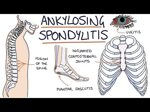
What Is Ankylosing Spondylitis (Axial Spondyloarthritis) and How Is It Treated?
Ankylosing Spondylitis (radiographic Axial Spondyloarthritis — axSpA) is a chronic HLA-B27–associated inflammatory rheumatic disease targeting the entheses (sites where ligaments, tendons, and joint capsules attach to bone) of the sacroiliac joints and axial skeleton. Chronic enthesitis undergoes ossification into bridging syndesmophytes ('bamboo spine'), leading to loss of lumbar lordosis, thoracic hyperkyphosis, costovertebral ankylosis (restrictive chest wall restriction), and hip flexion contractures. Crucially, daily spinal extension and deep costal expansion exercises are as fundamental as biologic/NSAID therapy!
Ankylosing spondylitis (inflammatory spinal rheumatism) is an inflammatory condition starting in the pelvic joints (sacroiliac joints) and spine in young adults. Unlike mechanical back pain (which feels better with rest), ankylosing spondylitis causes stiffness in the second half of the night and early morning (>45 minutes) that dramatically improves as soon as you exercise and take a warm shower!
Treatment and Exercise Videos
Phase-based physiotherapy protocols, mobilization, and therapeutic exercises
Step-by-step recovery and safe home exercise applications

Posture, Thoracic Expansion, and Spinal Mobility Exercises in Ankylosing Spondylitis
Phase 1: Acute Symptom Modulation, Tissue Protection & Early Activation: Phase 1 Clinical Focus & Tissue Protection: Daily Postural Extension & Prone Positioning Phase (Lifelong Core Habit): Mandatory 20-minute daily prone lying (flat on the stomach before sleep or in the morning) to prevent hip and thoracic flexion contractures, plus wall-referenced axial spinal extension (pressing heels, hips, shoulders, and occiput to the wall).
Step 1: Calming Pain, Protecting the Area & Gentle Starter Motion: Movement Is Your Medicine: In ankylosing spondylitis, resting makes the spine stiffer, while daily spinal extension and chest-opening exercises keep the vertebrae mobile and upright.

Hip and Spinal Mobility Exercises for Ankylosing Spondylitis and Rheumatoid Arthritis
Phase 2: Progressive Mobility, Motor Control & Targeted Strengthening: Phase 2 Progressive Mobilization & Motor Control: Costovertebral Thoracic Expansion Phase: Deep lateral/basal costal breathing with bilateral arm elevation (Thoracic Expansion Exercises) and scapular retraction to preserve chest expansion (>5 cm) and pulmonary vital capacity.
Step 2: Restoring Flexibility & Building Supporting Muscle Strength: The 20-Minute Tummy-Lying Habit: Lying flat on your stomach on a firm mattress for 20 minutes every day (and sleeping on a firm mattress with a very thin pillow) prevents stooped posture.
Clinical Assessment, Goniometry & Special Test Videos
Displayed only in Healthcare Professional mode (hidden in Patient mode)

Modified Schober Test — Clinical Measurement of Lumbar Spine Flexion Mobility
Goniometric ROM & Postural / Kinematic Analysis: BASMI (Bath Ankylosing Spondylitis Metrology Index)—1) Modified Schober Test (lumbar flexion <4–5 cm expansion indicates restriction), 2) Chest Expansion Difference at the 4th intercostal space (Normal: ≥5 cm; <2.5 cm indicates costovertebral ankylosis!), 3) Tragus-to-Wall / Occiput-to-Wall Distance (Normal: 0 cm), 4) Cervical Rotation, and 5) Intermalleolar Distance (hip abduction).
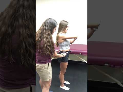
Vertebral Column (Cervical & Thoracolumbar Spine) Goniometric and Tape Measurement
Special Clinical Provocation & Neurological/Functional Tests: Sacroiliac provocation cluster (FABER, Gaenslen's, sacral thrust) and Thomas test (hip flexion contracture).

Sacroiliac Compression Test — Sacroiliac Joint Provocation Examination
Standardized Clinical Outcome Scales & Imaging Criteria: BASDAI (Disease Activity), BASFI (Functional Index), BASMI, and SIJ/spine MRI (active bone marrow edema sacroiliitis) & radiographs (New York criteria).