Multiple Sclerosis (MS)MS Disease (Multiple Sclerosis)
Multiple Sclerosis (MS) — Overview
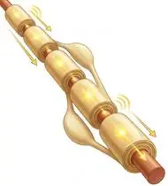
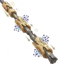
Multiple Sclerosis (MS) is an autoimmune inflammatory demyelinating and neurodegenerative disorder of the Central Nervous System (CNS) characterized by multifocal plaques of demyelination and axonal loss disseminated in space and time (Relapsing-Remitting RRMS ~85%, Secondary Progressive SPMS, Primary Progressive PPMS). Rehabilitation is governed by two cardinal neurophysiological rules: 1) Managing MS Fatigue & Energy Conservation (75–90% of patients experience central neurogenic fatigue) and 2) Preventing Uhthoff's Phenomenon (a rise in core body temperature of just 0.5°C transiently blocks nerve impulse conduction across demyelinated axons!).
Multiple Sclerosis (MS) is a condition in which the immune system creates small areas of inflammation on the protective insulating coating (myelin sheath) around nerve fibers in the brain and spinal cord. Regular, well-paced exercise is proven to boost brain growth factors (BDNF), improve balance, and reduce fatigue—as long as you stay cool and take rest breaks!
Primary Causes & Biomechanics
Key Points You Should Know
- •Autoimmune T-cell and B-cell destruction of CNS oligodendrocyte myelin sheaths slowing or blocking saltatory action potential conduction
- •Uhthoff's Phenomenon (Thermosensitivity): Heat (hot weather, hot showers, fever, or overheating during intense exercise) causes temporary conduction block and pseudo-exacerbation of visual/motor symptoms
- •Combined pyramidal spasticity/weakness (paraparesis/foot drop), cerebellar ataxia/tremor, and dorsal column proprioceptive sensory loss
- •Why Heat Temporarily Blurs Symptoms (Uhthoff's Rule): Warmth slows electrical signals across demyelinated nerves—so a hot room or overheating during exercise can make your vision or legs feel temporarily weaker. It is NOT new nerve damage and reverses as soon as you cool down! Always exercise in a cool room, sip cold water, and take cool showers.
- •The Energy Bank Account Rule: Never exercise to the point of total exhaustion. Leave energy in your tank so you feel refreshed 30–60 minutes after exercising.
- •Visual Focus for Balance (Frenkel Technique): Watching your foot and leg movements carefully with your eyes helps your brain guide smooth, steady steps.
Assessment Methods
1. Goniometric ROM & Postural / Kinematic Analysis
EDSS (Expanded Disability Status Scale 0–10), Modified Ashworth Scale (spasticity), and ankle dorsiflexion ROM.
2. Special Clinical Provocation & Neurological/Functional Tests
Modified Fatigue Impact Scale (MFIS), Berg Balance Scale (BBS), Timed 25-Foot Walk (T25FW), 6-Minute Walk Test, and Romberg / Frenkel coordination testing.
3. Standardized Clinical Outcome Scales & Imaging Criteria
EDSS, MSWS-12 (12-Item MS Walking Scale), MFIS, and brain/spinal cord MRI with gadolinium.
1. Range of Motion & Posture Check
Your physiotherapist gently evaluates how comfortably and smoothly your central nervous system white matter (oligodendrocyte myelin sheaths in optic nerves, periventricular brain, brainstem, cerebellum, and spinal cord) moves and checks your overall posture.
2. Muscle Strength & Functional Movement Check
Your muscle balance, walking or reaching comfort, and specific gentle movement tests are evaluated without triggering sharp pain.
3. Typical Symptoms & Daily Impact Review
Central Neurogenic Fatigue (most common symptom!) that worsens in the afternoon or with heat, accompanied by lower limb weakness, spasticity (leg stiffness/spasms), or fatigued foot drop (toes catching after walking 10–15 minutes).
Treatment Options & Phase-Based Protocol
✨ Enriched GIF Exercise & Precaution GuidePhase 1: Acute Symptom Modulation, Tissue Protection & Early Activation
- •Phase 1 Clinical Focus & Tissue Protection: Cool-Environment Spasticity & Energy Conservation Phase: Exercise strictly in a cool room (18–21°C) during rested morning hours with cold water/cooling vests & 2-minute rest intervals between sets! Perform slow towel gastro-soleus stretches, diaphragmatic breathing, and supine Frenkel precision heel slides.
- •Pain, Edema & Neuromuscular Modulation: Targeted physical modalities, relative load management, and early protected mobility of the central nervous system white matter (oligodendrocyte myelin sheaths in optic nerves, periventricular brain, brainstem, cerebellum, and spinal cord) without provoking mechanical or inflammatory irritation.
- •Proximal & Distal Kinetic Chain Maintenance: Maintain active mobility and circulation in adjacent joints while protecting the primary healing structures in Multiple Sclerosis.
Phase 2: Progressive Mobility, Motor Control & Targeted Strengthening
- •Phase 2 Progressive Mobilization & Motor Control: Submaximal Proximal & Core Stabilization Phase: Supine pelvic bridging, side-lying hip abduction, quadruped Bird-Dog, and seated terminal knee extension at a comfortable Borg RPE 11–13 (stopping well before exhaustion!).
- •Biomechanical Alignment & Co-Contraction: Restore balanced agonist-antagonist force couples, joint centration, and multi-planar neuromuscular control across the central nervous system white matter (oligodendrocyte myelin sheaths in optic nerves, periventricular brain, brainstem, cerebellum, and spinal cord).
- •Myofascial Extensibility & Endurance: Address secondary compensatory tightness and build local muscular endurance within safe, symptom-free biomechanical parameters.
Phase 3: Functional Integration, Proprioception & Long-Term Prevention
- •Phase 3 Functional Integration & Long-Term Prevention: Ataxia/Balance Integration & Functional Mobility Phase: Supported tandem/weight-shift balance training, chair sit-to-stand, ankle dorsiflexion / AFO or Functional Electrical Stimulation (FES) management for fatigued foot drop, and aquatic therapy in a cool pool (27–29°C, never a hot thermal pool!).
- •Proprioceptive Balance & Dynamic Loading: Progress to task-specific closed-chain, balance, and functional movement patterns tailored to daily life, occupational, or athletic demands.
- •Self-Management & Relapse Prophylaxis: Establish lifelong ergonomic habits, home exercise maintenance, and early warning-sign awareness to prevent recurrence.
Step 1: Calming Pain, Protecting the Area & Gentle Starter Motion
- •Why Heat Temporarily Blurs Symptoms (Uhthoff's Rule): Warmth slows electrical signals across demyelinated nerves—so a hot room or overheating during exercise can make your vision or legs feel temporarily weaker. It is NOT new nerve damage and reverses as soon as you cool down! Always exercise in a cool room, sip cold water, and take cool showers.
- •Calm Protection & Gentle Start: During the initial stage of MS Disease, avoid painful overloading and perform the gentle, pain-free starter movements shown below to support circulation and comfort.
- •Respecting Your Body's Signals: Every movement should stay within a comfortable, non-painful zone—never force through sharp or radiating pain.
Step 2: Restoring Flexibility & Building Supporting Muscle Strength
- •The Energy Bank Account Rule: Never exercise to the point of total exhaustion. Leave energy in your tank so you feel refreshed 30–60 minutes after exercising.
- •Step-by-Step Flexibility & Support: As acute sensitivity settles, gradually build mobility and strengthen the supporting muscles around the joint at a steady, controlled pace.
- •Posture & Breathing Coordination: Keep your posture upright and breathe smoothly (never hold your breath!) during every repetition.
Step 3: Safe Daily Habits, Stamina & Preventing Recurrence
- •Visual Focus for Balance (Frenkel Technique): Watching your foot and leg movements carefully with your eyes helps your brain guide smooth, steady steps.
- •Confident Return to Daily Life: Integrate your stronger muscles and improved balance into walking, stairs, work, and recreational activities.
- •Preventing Future Flare-Ups: Continue your core protective habits and key maintenance exercises even after symptoms resolve.
Animated (GIF) Therapeutic Exercise Guide
Animated clinical exercise protocol with biomechanical angles, target muscle groups, and dosage parameters (Click image to enlarge)
Step-by-step animated exercise demonstrations you can safely perform at home (Click image to enlarge)
 🔍 Enlarge
1. Supine Active-Assisted Heel Slide & Synovial Diffusion Mobilization
1. Supine Heel Slide (Gentle Knee & Hip Bending)
Controlled flexion and extension of the lower extremity by sliding the heel along the support surface without axial weight-bearing. Stimulates synovial fluid diffusion across avascular cartilage and prevents capsular adhesions. Specifically indicated in Multiple Sclerosis rehabilitation to optimize central nervous system white matter (oligodendrocyte myelin sheaths in optic nerves, periventricular brain, brainstem, cerebellum, and spinal cord) biomechanics and neuromuscular control.
While lying on your back, slowly slide your heel along the bed sheet toward your hip within a comfortable, pain-free angle, hold for 3 seconds, and gently slide it back out straight. Perform smoothly within a comfortable, pain-free range to support your MS Disease recovery.
 🔍 Enlarge
2. Long-Sitting Towel-Assisted Gastrocnemius-Soleus & Plantar Fascia Stretch
2. Seated Towel Calf & Foot Arch Stretch
Long-sitting with a towel looped around the metatarsal heads, applying a slow, sustained passive pull into ankle dorsiflexion. Restores gastrocnemius-soleus-Achilles length and reduces plantar fascial and equine tension. Specifically indicated in Multiple Sclerosis rehabilitation to optimize central nervous system white matter (oligodendrocyte myelin sheaths in optic nerves, periventricular brain, brainstem, cerebellum, and spinal cord) biomechanics and neuromuscular control.
Sit with your leg extended and loop a towel around the ball of your foot. Gently pull both ends of the towel toward you until you feel a comfortable stretch in your calf and sole, holding for 20–30 seconds. Perform smoothly within a comfortable, pain-free range to support your MS Disease recovery.
 🔍 Enlarge
3. Supine Lumbopelvic Bridging (Gluteus Maximus & Posterior Chain Co-Contraction)
3. Supine Hip Bridge (Controlled Pelvic Lift)
From hook-lying with transversus abdominis pre-activation, elevating the pelvis until hips, pelvis, and shoulders form a neutral diagonal line. Strengthens the gluteus maximus, hamstrings, and erector spinae synergy without lumbar hyperlordosis. Specifically indicated in Multiple Sclerosis rehabilitation to optimize central nervous system white matter (oligodendrocyte myelin sheaths in optic nerves, periventricular brain, brainstem, cerebellum, and spinal cord) biomechanics and neuromuscular control.
Lie on your back with your knees bent and feet flat hip-width apart. Gently tighten your tummy and glutes, lift your hips until your body forms a straight line from shoulders to knees, hold for 5 seconds, and lower slowly. Perform smoothly within a comfortable, pain-free range to support your MS Disease recovery.
 🔍 Enlarge
4. Quadruped Contralateral Arm-Leg Reach ('Bird-Dog' Multifidus & Deep Core)
4. All-Fours Opposite Arm & Leg Reach (Bird-Dog)
In quadruped with a neutral lumbopelvic spine and abdominal drawing-in (transversus abdominis), extending opposite upper and lower extremities horizontally without pelvic rotation or lumbar hyperextension. Minimizes spinal compressive load while maximizing multifidus recruitment. Specifically indicated in Multiple Sclerosis rehabilitation to optimize central nervous system white matter (oligodendrocyte myelin sheaths in optic nerves, periventricular brain, brainstem, cerebellum, and spinal cord) biomechanics and neuromuscular control.
On your hands and knees, keep your back flat like a tabletop and gently tighten your tummy. Slowly reach one arm forward and the opposite leg straight back withoutarching your lower back, hold for 5 seconds, and switch sides. Perform smoothly within a comfortable, pain-free range to support your MS Disease recovery.
 🔍 Enlarge
5. Functional Closed-Kinetic-Chain Chair Transfer (Sit-to-Stand Biomechanics)
5. Controlled Chair Sit-to-Stand Transfer
Controlled transition from sitting to standing and eccentric lowering back to the chair with symmetrical weight distribution, anterior trunk inclination ('nose over toes'), and neutral frontal knee alignment. Specifically indicated in Multiple Sclerosis rehabilitation to optimize central nervous system white matter (oligodendrocyte myelin sheaths in optic nerves, periventricular brain, brainstem, cerebellum, and spinal cord) biomechanics and neuromuscular control.
Sit near the front of a sturdy chair with your feet hip-width apart. Lean slightly forward and push evenly through both heels to stand up tall, then slowly lower yourself back down over 3 seconds. Perform smoothly within a comfortable, pain-free range to support your MS Disease recovery.
 🔍 Enlarge
6. Single-Leg / Semi-Tandem Proprioceptive & Weight-Shift Balance Training
6. Supported Weight-Shifting & Tandem Balance Practice
Controlled mediolateral/anteroposterior weight shifting and tandem or single-leg stance near a stable support. Enhances mechanoreceptor feedback, ankle/hip postural reflexes, and dynamic gait stability. Specifically indicated in Multiple Sclerosis rehabilitation to optimize central nervous system white matter (oligodendrocyte myelin sheaths in optic nerves, periventricular brain, brainstem, cerebellum, and spinal cord) biomechanics and neuromuscular control.
Stand near a counter or sturdy chair for safety. Place one foot slightly in front of the other (or shift your weight smoothly from side to side) and hold your balance upright for 20–30 seconds. Perform smoothly within a comfortable, pain-free range to support your MS Disease recovery.
Movements to Avoid & Strict Precautions
1. Exercising in Hot Rooms, Saunas, Turkish Baths (Hammam), or Hot Thermal Pools (>30°C!)
1. Using saunas, hot tubs, thermal spas, or exercising in hot/humid rooms (triggers Uhthoff's conduction block!)
2. Training to Complete Muscular Exhaustion Without Rest Intervals Between Sets
2. Pushing through exhaustion without taking 1–2 minute rest breaks between sets
3. Intense Exercise During an Active Acute MS Relapse or Fever
3. Doing heavy workouts during an active MS attack or while running a fever
4. Walking When Fatigued Without Using Recommended Mobility Aids (AFO / Cane) to Prevent Falls
4. Refusing to use a lightweight ankle brace or walking pole on long walks when your foot begins to drag
Multiple Sclerosis (MS) — Video Guide
Click directly on any video to open and watch in a window, or review details via 📋 Summary & Options.
Condition Overview Video (1 Video)
Summary of pathophysiology, biomechanics, and clinical presentation
What the condition is, why it develops, and its basic mechanism
What Is Multiple Sclerosis (MS)? Symptoms and Effects on the Nervous System
Multiple Sclerosis (MS) is an autoimmune inflammatory demyelinating and neurodegenerative disorder of the Central Nervous System (CNS) characterized by multifocal plaques of demyelination and axonal loss disseminated in space and time (Relapsing-Remitting RRMS ~85%, Secondary Progressive SPMS, Primary Progressive PPMS). Rehabilitation is governed by two cardinal neurophysiological rules: 1) Managing MS Fatigue & Energy Conservation (75–90% of patients experience central neurogenic fatigue) and 2) Preventing Uhthoff's Phenomenon (a rise in core body temperature of just 0.5°C transiently blocks nerve impulse conduction across demyelinated axons!).
Multiple Sclerosis (MS) is a condition in which the immune system creates small areas of inflammation on the protective insulating coating (myelin sheath) around nerve fibers in the brain and spinal cord. Regular, well-paced exercise is proven to boost brain growth factors (BDNF), improve balance, and reduce fatigue—as long as you stay cool and take rest breaks!
Treatment and Exercise Videos
Phase-based physiotherapy protocols, mobilization, and therapeutic exercises
Step-by-step recovery and safe home exercise applications

Balance and Coordination Exercises in Neurological Conditions (MS & Parkinson's)
Phase 1: Acute Symptom Modulation, Tissue Protection & Early Activation: Phase 1 Clinical Focus & Tissue Protection: Cool-Environment Spasticity & Energy Conservation Phase: Exercise strictly in a cool room (18–21°C) during rested morning hours with cold water/cooling vests & 2-minute rest intervals between sets! Perform slow towel gastro-soleus stretches, diaphragmatic breathing, and supine Frenkel precision heel slides.
Step 1: Calming Pain, Protecting the Area & Gentle Starter Motion: Why Heat Temporarily Blurs Symptoms (Uhthoff's Rule): Warmth slows electrical signals across demyelinated nerves—so a hot room or overheating during exercise can make your vision or legs feel temporarily weaker. It is NOT new nerve damage and reverses as soon as you cool down! Always exercise in a cool room, sip cold water, and take cool showers.

Static and Dynamic Balance Training in Neurological Rehabilitation
Phase 2: Progressive Mobility, Motor Control & Targeted Strengthening: Phase 2 Progressive Mobilization & Motor Control: Submaximal Proximal & Core Stabilization Phase: Supine pelvic bridging, side-lying hip abduction, quadruped Bird-Dog, and seated terminal knee extension at a comfortable Borg RPE 11–13 (stopping well before exhaustion!).
Step 2: Restoring Flexibility & Building Supporting Muscle Strength: The Energy Bank Account Rule: Never exercise to the point of total exhaustion. Leave energy in your tank so you feel refreshed 30–60 minutes after exercising.
Clinical Assessment, Goniometry & Special Test Videos
Displayed only in Healthcare Professional mode (hidden in Patient mode)

Systematic Neurological Examination: Reflex, Sensory, Tone, and Motor Assessment
Goniometric ROM & Postural / Kinematic Analysis: EDSS (Expanded Disability Status Scale 0–10), Modified Ashworth Scale (spasticity), and ankle dorsiflexion ROM.

Berg Balance Scale (BBS) — Clinical Postural Control and Balance Examination
Special Clinical Provocation & Neurological/Functional Tests: Modified Fatigue Impact Scale (MFIS), Berg Balance Scale (BBS), Timed 25-Foot Walk (T25FW), 6-Minute Walk Test, and Romberg / Frenkel coordination testing.

Modified Ashworth Scale (MAS) — Clinical Spasticity Assessment
Standardized Clinical Outcome Scales & Imaging Criteria: EDSS, MSWS-12 (12-Item MS Walking Scale), MFIS, and brain/spinal cord MRI with gadolinium.