Guillain-Barré Syndrome (GBS)Acute Peripheral Nerve Inflammation (Guillain-Barré Syndrome)
Guillain-Barré Syndrome (GBS) — Overview
Guillain-Barré Syndrome (GBS — most commonly Acute Inflammatory Demyelinating Polyradiculoneuropathy [AIDP]) is an acute, immune-mediated post-infectious polyradiculoneuropathy (often following *Campylobacter jejuni* gastroenteritis or respiratory viral infection by 1–3 weeks via molecular mimicry). It presents with rapidly progressive, symmetric ASCENDING flaccid paralysis (starting in the distal legs and ascending to the trunk, arms, cranial nerves VII/IX/X, and diaphragm, reaching its nadir within 2–4 weeks) accompanied by areflexia (loss of deep tendon reflexes). Following IVIG or plasmapheresis, 80–85% of patients achieve independent walking via phased remyelination and axonal regeneration—provided overwork fatigue (which causes paradoxical relapse of weakness!) and equinus/joint contractures are strictly prevented!
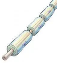
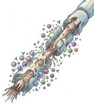
Guillain-Barré Syndrome (GBS / acute peripheral nerve inflammation) is a temporary condition—usually triggered 1 to 3 weeks after a stomach flu or cold—where the immune system mistakenly inflames the insulating coating (myelin) of the peripheral nerves outside the spinal cord. Unlike chronic nerve diseases, peripheral nerves regenerate their myelin coating, and the vast majority of GBS patients make a strong recovery with carefully paced rehabilitation!
Primary Causes & Biomechanics
Key Points You Should Know
- •Autoimmune antibody/macrophage attack on peripheral Schwann cell myelin sheaths or nodes of Ranvier causing acute conduction block
- •Three clinical phases: 1) Acute Progression Phase (Weeks 1–4, ICU/respiratory watch) -> 2) Plateau Phase (Days to Weeks) -> 3) Recovery / Remyelination Phase (Months 1–24)
- •Autonomic Dysfunction (in 65% of patients): labile blood pressure, orthostatic hypotension, and resting tachycardia/arrhythmias requiring heart-rate & BP monitoring during every therapy session!
- •Why Recovery Moves From Top to Bottom: Because nerve signals regrow from the spine outward toward the feet (about 1 mm per day), your hips, trunk, and shoulders get strong first, while your ankles and toes are the last to wake up—be patient and protect your ankles with splints while waiting!
- •The Overwork Rule (Why Doing Too Much Causes a Next-Day Dip): Newly remyelinating nerves tire out very quickly; if you exercise to exhaustion, your legs can feel weaker the next day. Always stop while you still feel fresh!
- •Calming Nerve Sensitivity: Warm water, loose cotton socks, and gentle slow stretching soothe the hypersensitive tingling skin sensation during nerve healing.
Assessment Methods
1. Goniometric ROM & Postural / Kinematic Analysis
MRC Sum Score (0–60 across 6 bilateral muscle groups), cranial nerve VII/bulbar screen, and ankle/hip/shoulder passive ROM.
2. Special Clinical Provocation & Neurological/Functional Tests
Forced Vital Capacity (FVC <20 mL/kg or rapid drop warrants ICU intubation readiness — '20/30/40 Rule'!), orthostatic blood pressure & heart rate monitoring, and neuropathic pain / dysesthesia VAS.
3. Standardized Clinical Outcome Scales & Imaging Criteria
GBS Disability Scale (Hughes 0–6), MRC Sum Score, Overall Neuropathy Limitation Scale (ONLS), and nerve conduction studies / CSF albuminocytologic dissociation.
1. Range of Motion & Posture Check
Your physiotherapist gently evaluates how comfortably and smoothly your peripheral nerve roots and peripheral nerves (Schwann cell myelin sheaths in AIDP, or nodal/axonal membranes in AMAN/AMSAN) moves and checks your overall posture.
2. Muscle Strength & Functional Movement Check
Your muscle balance, walking or reaching comfort, and specific gentle movement tests are evaluated without triggering sharp pain.
3. Typical Symptoms & Daily Impact Review
Rapidly Ascending Symmetric Weakness: Weakness and heaviness starting in both feet and legs (difficulty climbing stairs or rising from a chair) that spreads upward to the arms and sometimes facial muscles over days to 3 weeks.
Treatment Options & Phase-Based Protocol
✨ Enriched GIF Exercise & Precaution GuidePhase 1: Acute Symptom Modulation, Tissue Protection & Early Activation
- •Phase 1 Clinical Focus & Tissue Protection: Phase 1 — Acute / Plateau Phase (Contracture, DVT & Respiratory Protection): Strict avoidance of active fatigue! Perform gentle passive/active-assisted ankle pumps & towel calf stretches (preventing foot drop/equinus contractures with resting AFO splints!), supine heel slides, wand-assisted shoulder ROM, and diaphragmatic/costal breathing.
- •Pain, Edema & Neuromuscular Modulation: Targeted physical modalities, relative load management, and early protected mobility of the peripheral nerve roots and peripheral nerves (Schwann cell myelin sheaths in AIDP, or nodal/axonal membranes in AMAN/AMSAN) without provoking mechanical or inflammatory irritation.
- •Proximal & Distal Kinetic Chain Maintenance: Maintain active mobility and circulation in adjacent joints while protecting the primary healing structures in Guillain-Barré Syndrome.
Phase 2: Progressive Mobility, Motor Control & Targeted Strengthening
- •Phase 2 Progressive Mobilization & Motor Control: Phase 2 — Early Remyelination & Gravity-Reduced Phase: Gradual upright tilt-table/bed sitting (monitoring orthostatic hypotension!), submaximal isometric quad sets, pelvic bridging, and active finger/wrist mobility with 2-minute full rest breaks between sets.
- •Biomechanical Alignment & Co-Contraction: Restore balanced agonist-antagonist force couples, joint centration, and multi-planar neuromuscular control across the peripheral nerve roots and peripheral nerves (Schwann cell myelin sheaths in AIDP, or nodal/axonal membranes in AMAN/AMSAN).
- •Myofascial Extensibility & Endurance: Address secondary compensatory tightness and build local muscular endurance within safe, symptom-free biomechanical parameters.
Phase 3: Functional Integration, Proprioception & Long-Term Prevention
- •Phase 3 Functional Integration & Long-Term Prevention: Phase 3 — Proximal-to-Distal Reinnervation & Ambulation Phase: Side-lying/standing hip abduction, chair sit-to-stand, supported tandem balance, and gait training (using temporary AFOs until distal tibialis anterior reinnervation completes!).
- •Proprioceptive Balance & Dynamic Loading: Progress to task-specific closed-chain, balance, and functional movement patterns tailored to daily life, occupational, or athletic demands.
- •Self-Management & Relapse Prophylaxis: Establish lifelong ergonomic habits, home exercise maintenance, and early warning-sign awareness to prevent recurrence.
Step 1: Calming Pain, Protecting the Area & Gentle Starter Motion
- •Why Recovery Moves From Top to Bottom: Because nerve signals regrow from the spine outward toward the feet (about 1 mm per day), your hips, trunk, and shoulders get strong first, while your ankles and toes are the last to wake up—be patient and protect your ankles with splints while waiting!
- •Calm Protection & Gentle Start: During the initial stage of Acute Peripheral Nerve Inflammation, avoid painful overloading and perform the gentle, pain-free starter movements shown below to support circulation and comfort.
- •Respecting Your Body's Signals: Every movement should stay within a comfortable, non-painful zone—never force through sharp or radiating pain.
Step 2: Restoring Flexibility & Building Supporting Muscle Strength
- •The Overwork Rule (Why Doing Too Much Causes a Next-Day Dip): Newly remyelinating nerves tire out very quickly; if you exercise to exhaustion, your legs can feel weaker the next day. Always stop while you still feel fresh!
- •Step-by-Step Flexibility & Support: As acute sensitivity settles, gradually build mobility and strengthen the supporting muscles around the joint at a steady, controlled pace.
- •Posture & Breathing Coordination: Keep your posture upright and breathe smoothly (never hold your breath!) during every repetition.
Step 3: Safe Daily Habits, Stamina & Preventing Recurrence
- •Calming Nerve Sensitivity: Warm water, loose cotton socks, and gentle slow stretching soothe the hypersensitive tingling skin sensation during nerve healing.
- •Confident Return to Daily Life: Integrate your stronger muscles and improved balance into walking, stairs, work, and recreational activities.
- •Preventing Future Flare-Ups: Continue your core protective habits and key maintenance exercises even after symptoms resolve.
Animated (GIF) Therapeutic Exercise Guide
Animated clinical exercise protocol with biomechanical angles, target muscle groups, and dosage parameters (Click image to enlarge)
Step-by-step animated exercise demonstrations you can safely perform at home (Click image to enlarge)
 🔍 Enlarge
1. Rhythmic Active Ankle Dorsiflexion & Plantarflexion Pumping (Venous/Edema Pump)
1. Ankle Toe-Pulls and Pointing (Circulation & Swelling Pump)
Rhythmic alternating contraction of the tibialis anterior and gastrocnemius-soleus complex. Activates the deep venous calf pump to prevent deep vein thrombosis (DVT), clears distal edema, and maintains talocrural mobility. Specifically indicated in Guillain-Barré Syndrome rehabilitation to optimize peripheral nerve roots and peripheral nerves (Schwann cell myelin sheaths in AIDP, or nodal/axonal membranes in AMAN/AMSAN) biomechanics and neuromuscular control.
While lying down or sitting with your legs supported, pull your toes and feet firmly upward toward your shins, hold for 3 seconds, then point them downward like pressing a gas pedal. Perform smoothly within a comfortable, pain-free range to support your Acute Peripheral Nerve Inflammation recovery.
 🔍 Enlarge
2. Supine Active-Assisted Heel Slide & Synovial Diffusion Mobilization
2. Supine Heel Slide (Gentle Knee & Hip Bending)
Controlled flexion and extension of the lower extremity by sliding the heel along the support surface without axial weight-bearing. Stimulates synovial fluid diffusion across avascular cartilage and prevents capsular adhesions. Specifically indicated in Guillain-Barré Syndrome rehabilitation to optimize peripheral nerve roots and peripheral nerves (Schwann cell myelin sheaths in AIDP, or nodal/axonal membranes in AMAN/AMSAN) biomechanics and neuromuscular control.
While lying on your back, slowly slide your heel along the bed sheet toward your hip within a comfortable, pain-free angle, hold for 3 seconds, and gently slide it back out straight. Perform smoothly within a comfortable, pain-free range to support your Acute Peripheral Nerve Inflammation recovery.
 🔍 Enlarge
3. Isometric Quadriceps Setting (Popliteal Towel Press)
3. Under-Knee Towel Press (Quadriceps Tightening)
Isometric compression of a rolled towel beneath the popliteal fossa into the surface with simultaneous ankle dorsiflexion. Activates the Vastus Medialis Obliquus (VMO) and overcomes arthrogenic muscle inhibition with 0° joint shear stress. Specifically indicated in Guillain-Barré Syndrome rehabilitation to optimize peripheral nerve roots and peripheral nerves (Schwann cell myelin sheaths in AIDP, or nodal/axonal membranes in AMAN/AMSAN) biomechanics and neuromuscular control.
Place a small rolled towel under your knee while lying on your back or sitting with your leg straight. Pull your toes toward you while firmly pressing the back of your knee down into the towel, hold for 5 seconds, and relax while exhaling. Perform smoothly within a comfortable, pain-free range to support your Acute Peripheral Nerve Inflammation recovery.
 🔍 Enlarge
4. Supine Lumbopelvic Bridging (Gluteus Maximus & Posterior Chain Co-Contraction)
4. Supine Hip Bridge (Controlled Pelvic Lift)
From hook-lying with transversus abdominis pre-activation, elevating the pelvis until hips, pelvis, and shoulders form a neutral diagonal line. Strengthens the gluteus maximus, hamstrings, and erector spinae synergy without lumbar hyperlordosis. Specifically indicated in Guillain-Barré Syndrome rehabilitation to optimize peripheral nerve roots and peripheral nerves (Schwann cell myelin sheaths in AIDP, or nodal/axonal membranes in AMAN/AMSAN) biomechanics and neuromuscular control.
Lie on your back with your knees bent and feet flat hip-width apart. Gently tighten your tummy and glutes, lift your hips until your body forms a straight line from shoulders to knees, hold for 5 seconds, and lower slowly. Perform smoothly within a comfortable, pain-free range to support your Acute Peripheral Nerve Inflammation recovery.
 🔍 Enlarge
5. Functional Closed-Kinetic-Chain Chair Transfer (Sit-to-Stand Biomechanics)
5. Controlled Chair Sit-to-Stand Transfer
Controlled transition from sitting to standing and eccentric lowering back to the chair with symmetrical weight distribution, anterior trunk inclination ('nose over toes'), and neutral frontal knee alignment. Specifically indicated in Guillain-Barré Syndrome rehabilitation to optimize peripheral nerve roots and peripheral nerves (Schwann cell myelin sheaths in AIDP, or nodal/axonal membranes in AMAN/AMSAN) biomechanics and neuromuscular control.
Sit near the front of a sturdy chair with your feet hip-width apart. Lean slightly forward and push evenly through both heels to stand up tall, then slowly lower yourself back down over 3 seconds. Perform smoothly within a comfortable, pain-free range to support your Acute Peripheral Nerve Inflammation recovery.
 🔍 Enlarge
6. Single-Leg / Semi-Tandem Proprioceptive & Weight-Shift Balance Training
6. Supported Weight-Shifting & Tandem Balance Practice
Controlled mediolateral/anteroposterior weight shifting and tandem or single-leg stance near a stable support. Enhances mechanoreceptor feedback, ankle/hip postural reflexes, and dynamic gait stability. Specifically indicated in Guillain-Barré Syndrome rehabilitation to optimize peripheral nerve roots and peripheral nerves (Schwann cell myelin sheaths in AIDP, or nodal/axonal membranes in AMAN/AMSAN) biomechanics and neuromuscular control.
Stand near a counter or sturdy chair for safety. Place one foot slightly in front of the other (or shift your weight smoothly from side to side) and hold your balance upright for 20–30 seconds. Perform smoothly within a comfortable, pain-free range to support your Acute Peripheral Nerve Inflammation recovery.
Movements to Avoid & Strict Precautions
1. Exercising to Muscular Exhaustion or Failure (Causes Demyelinated Conduction Block & Next-Day Weakness!)
1. Pushing your muscles to exhaustion or until they tremble (always stop with energy in reserve and rest 2 minutes between sets!)
2. Letting the Feet Drop Pointed Downward Under Heavy Bed Covers (Causes Permanent Equinus Contracture!)
2. Letting heavy blankets push your feet downward in bed without resting ankle splints/pillows
3. Standing Up Suddenly From Lying Down (Triggers Severe Orthostatic Blood Pressure Drop!)
3. Standing up quickly without sitting at the edge of the bed for 1–2 minutes first
4. Walking Barefoot With a Dragging Foot Drop (High Risk of Ankle Sprain & Falls!)
4. Walking without your supportive ankle brace (AFO) while the foot-lifting muscles are still recovering
Guillain-Barré Syndrome (GBS) — Video Guide
Click directly on any video to open and watch in a window, or review details via 📋 Summary & Options.
Condition Overview Video (1 Video)
Summary of pathophysiology, biomechanics, and clinical presentation
What the condition is, why it develops, and its basic mechanism

What Is Guillain-Barré Syndrome (GBS / Acute Polyneuropathy)? Course and Treatment
Guillain-Barré Syndrome (GBS — most commonly Acute Inflammatory Demyelinating Polyradiculoneuropathy [AIDP]) is an acute, immune-mediated post-infectious polyradiculoneuropathy (often following *Campylobacter jejuni* gastroenteritis or respiratory viral infection by 1–3 weeks via molecular mimicry). It presents with rapidly progressive, symmetric ASCENDING flaccid paralysis (starting in the distal legs and ascending to the trunk, arms, cranial nerves VII/IX/X, and diaphragm, reaching its nadir within 2–4 weeks) accompanied by areflexia (loss of deep tendon reflexes). Following IVIG or plasmapheresis, 80–85% of patients achieve independent walking via phased remyelination and axonal regeneration—provided overwork fatigue (which causes paradoxical relapse of weakness!) and equinus/joint contractures are strictly prevented!
Guillain-Barré Syndrome (GBS / acute peripheral nerve inflammation) is a temporary condition—usually triggered 1 to 3 weeks after a stomach flu or cold—where the immune system mistakenly inflames the insulating coating (myelin) of the peripheral nerves outside the spinal cord. Unlike chronic nerve diseases, peripheral nerves regenerate their myelin coating, and the vast majority of GBS patients make a strong recovery with carefully paced rehabilitation!
Treatment and Exercise Videos
Phase-based physiotherapy protocols, mobilization, and therapeutic exercises
Step-by-step recovery and safe home exercise applications

Ankle Strengthening and Stretching Exercises in Polyneuropathy and Neuro Rehab
Phase 1: Acute Symptom Modulation, Tissue Protection & Early Activation: Phase 1 Clinical Focus & Tissue Protection: Phase 1 — Acute / Plateau Phase (Contracture, DVT & Respiratory Protection): Strict avoidance of active fatigue! Perform gentle passive/active-assisted ankle pumps & towel calf stretches (preventing foot drop/equinus contractures with resting AFO splints!), supine heel slides, wand-assisted shoulder ROM, and diaphragmatic/costal breathing.
Step 1: Calming Pain, Protecting the Area & Gentle Starter Motion: Why Recovery Moves From Top to Bottom: Because nerve signals regrow from the spine outward toward the feet (about 1 mm per day), your hips, trunk, and shoulders get strong first, while your ankles and toes are the last to wake up—be patient and protect your ankles with splints while waiting!

Static and Dynamic Balance Training in Neurological Rehabilitation
Phase 2: Progressive Mobility, Motor Control & Targeted Strengthening: Phase 2 Progressive Mobilization & Motor Control: Phase 2 — Early Remyelination & Gravity-Reduced Phase: Gradual upright tilt-table/bed sitting (monitoring orthostatic hypotension!), submaximal isometric quad sets, pelvic bridging, and active finger/wrist mobility with 2-minute full rest breaks between sets.
Step 2: Restoring Flexibility & Building Supporting Muscle Strength: The Overwork Rule (Why Doing Too Much Causes a Next-Day Dip): Newly remyelinating nerves tire out very quickly; if you exercise to exhaustion, your legs can feel weaker the next day. Always stop while you still feel fresh!
Clinical Assessment, Goniometry & Special Test Videos
Displayed only in Healthcare Professional mode (hidden in Patient mode)

Systematic Neurological Examination: Reflex, Sensory, Tone, and Motor Assessment
Goniometric ROM & Postural / Kinematic Analysis: MRC Sum Score (0–60 across 6 bilateral muscle groups), cranial nerve VII/bulbar screen, and ankle/hip/shoulder passive ROM.

Manual Muscle Testing (MRC 0–5 Grading) — Clinical Motor Strength Examination
Special Clinical Provocation & Neurological/Functional Tests: Forced Vital Capacity (FVC <20 mL/kg or rapid drop warrants ICU intubation readiness — '20/30/40 Rule'!), orthostatic blood pressure & heart rate monitoring, and neuropathic pain / dysesthesia VAS.