Duchenne Muscular Dystrophy (DMD)Progressive Muscle Wasting Disease (Duchenne Muscular Dystrophy)
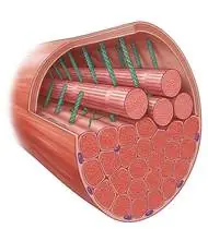
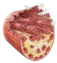
Duchenne Muscular Dystrophy (DMD) — Overview
Duchenne Muscular Dystrophy (DMD) is the most common and severe X-linked recessive muscular dystrophy, caused by frameshift mutations in the DMD gene (Xp21) resulting in complete absence of functional dystrophin protein at the muscle fiber membrane (sarcolemma). Without dystrophin to anchor the actin cytoskeleton to the extracellular matrix, mechanical contraction—especially ECCENTRIC contraction—shears the fragile sarcolemma, allowing lethal calcium influx, myofiber necrosis, and replacement by fat and fibrous tissue (calf pseudohypertrophy). GOLD STANDARD PHYSIOTHERAPY RULE: Eccentric and high-resistance exercises are STRICTLY CONTRAINDICATED because they mechanically tear the dystrophin-deficient sarcolemma!
Duchenne Muscular Dystrophy (DMD) is a genetic muscle condition in which the body lacks dystrophin—the shock-absorbing protein that protects muscle cell walls during movement. Because muscle cells without dystrophin are delicate, physiotherapy in DMD is built on two life-changing pillars: 1) Daily gentle stretching + night splints + pool exercise (which can extend independent walking by 2 to 4 years!) and 2) Avoiding heavy or lowering ('eccentric') loads that damage muscle cells.
Primary Causes & Biomechanics
Key Points You Should Know
- •Absence of sarcolemmal dystrophin causing mechanical membrane fragility, chronic inflammation, and fibro-fatty pseudohypertrophy
- •Predictable Contracture Cascade: 1) Gastrocnemius-Soleus (Equinus / toe-walking) -> 2) Iliotibial Band (ITB) & Hip Flexors (Iliopsoas/TFL) -> 3) Knee Flexors (Hamstrings) -> 4) Neuromuscular Scoliosis & Wrist/Finger Flexors
- •Progressive dilated cardiomyopathy and restrictive respiratory insufficiency in adolescence/adulthood
- •Why Eccentric (Lowering) Exercises Are Strictly Forbidden: Lowering a heavy weight, jumping down, or walking down steep stairs stretches a contracting muscle and tears the delicate dystrophin-free cell wall! Always stick to gentle, non-fatiguing movements and swimming/pool play.
- •Evening Calf Stretches + Night Splints (AFOs): The Achilles tendon is always the first to tighten in DMD (causing toe-walking). Stretching the calves gently every evening after a warm bath and wearing resting ankle splints at night in bed keeps the feet flat and preserves walking for years longer!
- •Daily 20-Minute Tummy Time: Lying flat on the stomach while reading or watching a tablet stretches the front of the hips and stops the lower back from arching excessively.
Assessment Methods
1. Goniometric ROM & Postural / Kinematic Analysis
North Star Ambulatory Assessment (NSAA, 0–34 points), Timed Function Tests (Time to Stand from Supine [Gowers' maneuver], 10-Meter Walk/Run, 4-Stair Climb), PUL (Performance of Upper Limb), and ankle dorsiflexion (<0° indicates equinus contracture!) / Thomas & Ober tests.
2. Special Clinical Provocation & Neurological/Functional Tests
Gowers' Sign (the patient rolls prone, pushes up on hands and feet, and 'walks' the hands up the shins and thighs to extend the weak gluteus maximus and quadriceps!), Forced Vital Capacity (FVC), and Peak Cough Flow (PCF).
3. Standardized Clinical Outcome Scales & Imaging Criteria
NSAA, Timed Gowers' Stand (<5s vs. >10s predicts time to loss of ambulation), 6MWT, PUL 2.0, and FVC / Echocardiogram.
1. Range of Motion & Posture Check
Your physiotherapist gently evaluates how comfortably and smoothly your striated skeletal and cardiac muscle sarcolemma (X-linked absence of dystrophin protein — Xp21 DMD gene) moves and checks your overall posture.
2. Muscle Strength & Functional Movement Check
Your muscle balance, walking or reaching comfort, and specific gentle movement tests are evaluated without triggering sharp pain.
3. Typical Symptoms & Daily Impact Review
Proximal Pelvic & Thigh Weakness: Difficulty rising from the floor (Gowers' sign—using the hands to 'climb up' the thighs), waddling gait (Trendelenburg), difficulty climbing stairs, and frequent falls.
Treatment Options & Phase-Based Protocol
✨ Enriched GIF Exercise & Precaution GuidePhase 1: Acute Symptom Modulation, Tissue Protection & Early Activation
- •Phase 1 Clinical Focus & Tissue Protection: Daily Contracture Delay & Night Splinting Phase (THE #1 PRIORITY IN DMD!): Daily slow, pain-free passive/active-assisted stretching (3x30–60s every evening) of the gastrocnemius-soleus (towel stretch), iliotibial band, hip flexors (20 min prone lying!), and hamstrings, combined with mandatory Resting Night AFOs (never dynamic daytime AFOs while walking, as DMD patients rely on slight equinus to create a knee extension moment!).
- •Pain, Edema & Neuromuscular Modulation: Targeted physical modalities, relative load management, and early protected mobility of the striated skeletal and cardiac muscle sarcolemma (X-linked absence of dystrophin protein — Xp21 DMD gene) without provoking mechanical or inflammatory irritation.
- •Proximal & Distal Kinetic Chain Maintenance: Maintain active mobility and circulation in adjacent joints while protecting the primary healing structures in Duchenne Muscular Dystrophy.
Phase 2: Progressive Mobility, Motor Control & Targeted Strengthening
- •Phase 2 Progressive Mobilization & Motor Control: Submaximal Concentric / Aquatic & Postural Phase: Strictly NO eccentric or heavy weights! Perform gentle submaximal concentric/active movements, aquatic therapy in a warm pool (buoyancy allows joyful movement with zero sarcolemmal tear!), unloaded supine heel slides, and scapular retraction.
- •Biomechanical Alignment & Co-Contraction: Restore balanced agonist-antagonist force couples, joint centration, and multi-planar neuromuscular control across the striated skeletal and cardiac muscle sarcolemma (X-linked absence of dystrophin protein — Xp21 DMD gene).
- •Myofascial Extensibility & Endurance: Address secondary compensatory tightness and build local muscular endurance within safe, symptom-free biomechanical parameters.
Phase 3: Functional Integration, Proprioception & Long-Term Prevention
- •Phase 3 Functional Integration & Long-Term Prevention: Respiratory, Scoliosis & Upper Limb Independence Phase: Diaphragmatic & thoracic expansion breathing, breath-stacking & Cough Assist, standing frame / KAFO prolonged standing (delays scoliosis and preserves bone density!), and hand/wrist tendon gliding & thumb opposition.
- •Proprioceptive Balance & Dynamic Loading: Progress to task-specific closed-chain, balance, and functional movement patterns tailored to daily life, occupational, or athletic demands.
- •Self-Management & Relapse Prophylaxis: Establish lifelong ergonomic habits, home exercise maintenance, and early warning-sign awareness to prevent recurrence.
Step 1: Calming Pain, Protecting the Area & Gentle Starter Motion
- •Why Eccentric (Lowering) Exercises Are Strictly Forbidden: Lowering a heavy weight, jumping down, or walking down steep stairs stretches a contracting muscle and tears the delicate dystrophin-free cell wall! Always stick to gentle, non-fatiguing movements and swimming/pool play.
- •Calm Protection & Gentle Start: During the initial stage of Progressive Muscle Wasting Disease, avoid painful overloading and perform the gentle, pain-free starter movements shown below to support circulation and comfort.
- •Respecting Your Body's Signals: Every movement should stay within a comfortable, non-painful zone—never force through sharp or radiating pain.
Step 2: Restoring Flexibility & Building Supporting Muscle Strength
- •Evening Calf Stretches + Night Splints (AFOs): The Achilles tendon is always the first to tighten in DMD (causing toe-walking). Stretching the calves gently every evening after a warm bath and wearing resting ankle splints at night in bed keeps the feet flat and preserves walking for years longer!
- •Step-by-Step Flexibility & Support: As acute sensitivity settles, gradually build mobility and strengthen the supporting muscles around the joint at a steady, controlled pace.
- •Posture & Breathing Coordination: Keep your posture upright and breathe smoothly (never hold your breath!) during every repetition.
Step 3: Safe Daily Habits, Stamina & Preventing Recurrence
- •Daily 20-Minute Tummy Time: Lying flat on the stomach while reading or watching a tablet stretches the front of the hips and stops the lower back from arching excessively.
- •Confident Return to Daily Life: Integrate your stronger muscles and improved balance into walking, stairs, work, and recreational activities.
- •Preventing Future Flare-Ups: Continue your core protective habits and key maintenance exercises even after symptoms resolve.
Animated (GIF) Therapeutic Exercise Guide
Animated clinical exercise protocol with biomechanical angles, target muscle groups, and dosage parameters (Click image to enlarge)
Step-by-step animated exercise demonstrations you can safely perform at home (Click image to enlarge)
 🔍 Enlarge
1. Long-Sitting Towel-Assisted Gastrocnemius-Soleus & Plantar Fascia Stretch
1. Seated Towel Calf & Foot Arch Stretch
Long-sitting with a towel looped around the metatarsal heads, applying a slow, sustained passive pull into ankle dorsiflexion. Restores gastrocnemius-soleus-Achilles length and reduces plantar fascial and equine tension. Specifically indicated in Duchenne Muscular Dystrophy rehabilitation to optimize striated skeletal and cardiac muscle sarcolemma (X-linked absence of dystrophin protein — Xp21 DMD gene) biomechanics and neuromuscular control.
Sit with your leg extended and loop a towel around the ball of your foot. Gently pull both ends of the towel toward you until you feel a comfortable stretch in your calf and sole, holding for 20–30 seconds. Perform smoothly within a comfortable, pain-free range to support your Progressive Muscle Wasting Disease recovery.
 🔍 Enlarge
2. Supine Active-Assisted Heel Slide & Synovial Diffusion Mobilization
2. Supine Heel Slide (Gentle Knee & Hip Bending)
Controlled flexion and extension of the lower extremity by sliding the heel along the support surface without axial weight-bearing. Stimulates synovial fluid diffusion across avascular cartilage and prevents capsular adhesions. Specifically indicated in Duchenne Muscular Dystrophy rehabilitation to optimize striated skeletal and cardiac muscle sarcolemma (X-linked absence of dystrophin protein — Xp21 DMD gene) biomechanics and neuromuscular control.
While lying on your back, slowly slide your heel along the bed sheet toward your hip within a comfortable, pain-free angle, hold for 3 seconds, and gently slide it back out straight. Perform smoothly within a comfortable, pain-free range to support your Progressive Muscle Wasting Disease recovery.
 🔍 Enlarge
3. Dorsal/Palmar Interossei & Lumbrical Activation (Active Finger Abduction/Extension)
3. Tabletop Finger Spreading (Fan Out) & Opening
With the palm resting flat on a surface, actively spreading all digits apart (abduction) and bringing them back together, combined with controlled MCP/IP extension. Strengthens ulnar-innervated interossei and balances extrinsic flexors. Specifically indicated in Duchenne Muscular Dystrophy rehabilitation to optimize striated skeletal and cardiac muscle sarcolemma (X-linked absence of dystrophin protein — Xp21 DMD gene) biomechanics and neuromuscular control.
Rest your hand flat on a table. Spread your fingers wide apart like a fan, hold for 3 seconds, and slowly bring them back together without lifting your palm. Perform smoothly within a comfortable, pain-free range to support your Progressive Muscle Wasting Disease recovery.
 🔍 Enlarge
4. Diaphragmatic & Pursed-Lip Breathing (PLB — Autonomic & Ventilatory Control)
4. Calm Belly Breathing with Pursed-Lip Exhale
Slow nasal inhalation expanding the lower ribcage and abdomen (while relaxing accessory neck/shoulder muscles), followed by prolonged expiration through pursed lips at a 1:2 ratio. Prevents dynamic airway collapse, improves basal ventilation, and reduces sympathetic tone. Specifically indicated in Duchenne Muscular Dystrophy rehabilitation to optimize striated skeletal and cardiac muscle sarcolemma (X-linked absence of dystrophin protein — Xp21 DMD gene) biomechanics and neuromuscular control.
Sit comfortably and place one hand on your chest and the other on your belly. Breathe in slowly through your nose for 2–3 seconds so your belly gently rises, then breathe out slowly for 4–6 seconds through pursed lips as if flickering a candle. Perform smoothly within a comfortable, pain-free range to support your Progressive Muscle Wasting Disease recovery.
 🔍 Enlarge
5. Scapulothoracic Retraction & Depression (Middle/Lower Trapezius & Rhomboids)
5. Shoulder Blade Squeeze (Postural Alignment)
Adduction and depression of the scapulae via the middle/lower trapezius and rhomboids while inhibiting upper trapezius overactivity. Corrects anterior scapular tilt, widens the subacromial outlet, and unloads the anterior thoracic column. Specifically indicated in Duchenne Muscular Dystrophy rehabilitation to optimize striated skeletal and cardiac muscle sarcolemma (X-linked absence of dystrophin protein — Xp21 DMD gene) biomechanics and neuromuscular control.
Sit or stand tall. Keep your shoulders relaxed away from your ears, gently draw your shoulder blades together and slightly downward toward your back pockets, hold for 5 seconds, and release. Perform smoothly within a comfortable, pain-free range to support your Progressive Muscle Wasting Disease recovery.
 🔍 Enlarge
6. Prone Positioning & Controlled Thoracolumbar Extension (McKenzie / Back Extensor)
6. Prone Lying & Gentle Back Extension Lift
Prone positioning and graduated thoracic/lumbar extension (with a pillow under the abdomen when indicated for spinal unloading). Counters flexed postural kyphosis, stretches tight hip flexors, and strengthens paraspinal extensors. Specifically indicated in Duchenne Muscular Dystrophy rehabilitation to optimize striated skeletal and cardiac muscle sarcolemma (X-linked absence of dystrophin protein — Xp21 DMD gene) biomechanics and neuromuscular control.
Lie on your stomach (place a small pillow under your hips/tummy if needed for comfort). Gently lift your head and chest slightly off the mat by squeezing your back muscles without straining your neck, hold for 3–5 seconds, and lower. Perform smoothly within a comfortable, pain-free range to support your Progressive Muscle Wasting Disease recovery.
Movements to Avoid & Strict Precautions
1. Eccentric Exercises, Heavy Weights, Downhill Running, Trampolines, or Jumping Down From Heights (Tears Muscle Cell Membranes!)
1. Jumping on trampolines, jumping down from steps, walking down long flights of stairs, or lifting weights (STRICTLY FORBIDDEN!)
2. Wearing Rigid Ankle Braces (AFOs) During Daytime Walking (Blocks the Compensatory Knee Lock and Causes Falls!)
2. Wearing stiff ankle splints while walking during the day (AFO splints in DMD are worn AT NIGHT in bed or while sitting, unless specifically guided by your specialist!)
3. Skipping Daily Evening Calf & Hip Flexor Stretches
3. Skipping evening calf stretches and tummy-lying hip stretches
4. Prolonged Immobilization After a Minor Injury or Fracture (Causes Rapid Disuse Loss of Walking!)
4. Staying immobilized in bed for weeks after a minor sprain or fracture (early supported standing is critical!)
Duchenne Muscular Dystrophy (DMD) — Video Guide
Click directly on any video to open and watch in a window, or review details via 📋 Summary & Options.
Condition Overview Video (1 Video)
Summary of pathophysiology, biomechanics, and clinical presentation
What the condition is, why it develops, and its basic mechanism

What Are Duchenne and Becker Muscular Dystrophy (DMD / BMD)?
Duchenne Muscular Dystrophy (DMD) is the most common and severe X-linked recessive muscular dystrophy, caused by frameshift mutations in the DMD gene (Xp21) resulting in complete absence of functional dystrophin protein at the muscle fiber membrane (sarcolemma). Without dystrophin to anchor the actin cytoskeleton to the extracellular matrix, mechanical contraction—especially ECCENTRIC contraction—shears the fragile sarcolemma, allowing lethal calcium influx, myofiber necrosis, and replacement by fat and fibrous tissue (calf pseudohypertrophy). GOLD STANDARD PHYSIOTHERAPY RULE: Eccentric and high-resistance exercises are STRICTLY CONTRAINDICATED because they mechanically tear the dystrophin-deficient sarcolemma!
Duchenne Muscular Dystrophy (DMD) is a genetic muscle condition in which the body lacks dystrophin—the shock-absorbing protein that protects muscle cell walls during movement. Because muscle cells without dystrophin are delicate, physiotherapy in DMD is built on two life-changing pillars: 1) Daily gentle stretching + night splints + pool exercise (which can extend independent walking by 2 to 4 years!) and 2) Avoiding heavy or lowering ('eccentric') loads that damage muscle cells.
Treatment and Exercise Videos
Phase-based physiotherapy protocols, mobilization, and therapeutic exercises
Step-by-step recovery and safe home exercise applications

Physiotherapy, Submaximal Exercise, and Contracture Management in Muscular Dystrophies
Phase 1: Acute Symptom Modulation, Tissue Protection & Early Activation: Phase 1 Clinical Focus & Tissue Protection: Daily Contracture Delay & Night Splinting Phase (THE #1 PRIORITY IN DMD!): Daily slow, pain-free passive/active-assisted stretching (3x30–60s every evening) of the gastrocnemius-soleus (towel stretch), iliotibial band, hip flexors (20 min prone lying!), and hamstrings, combined with mandatory Resting Night AFOs (never dynamic daytime AFOs while walking, as DMD patients rely on slight equinus to create a knee extension moment!).
Step 1: Calming Pain, Protecting the Area & Gentle Starter Motion: Why Eccentric (Lowering) Exercises Are Strictly Forbidden: Lowering a heavy weight, jumping down, or walking down steep stairs stretches a contracting muscle and tears the delicate dystrophin-free cell wall! Always stick to gentle, non-fatiguing movements and swimming/pool play.
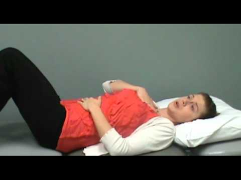
Diaphragmatic Breathing (Abdominal Breathing) Instruction and Clinical Application
Phase 2: Progressive Mobility, Motor Control & Targeted Strengthening: Phase 2 Progressive Mobilization & Motor Control: Submaximal Concentric / Aquatic & Postural Phase: Strictly NO eccentric or heavy weights! Perform gentle submaximal concentric/active movements, aquatic therapy in a warm pool (buoyancy allows joyful movement with zero sarcolemmal tear!), unloaded supine heel slides, and scapular retraction.
Step 2: Restoring Flexibility & Building Supporting Muscle Strength: Evening Calf Stretches + Night Splints (AFOs): The Achilles tendon is always the first to tighten in DMD (causing toe-walking). Stretching the calves gently every evening after a warm bath and wearing resting ankle splints at night in bed keeps the feet flat and preserves walking for years longer!
Clinical Assessment, Goniometry & Special Test Videos
Displayed only in Healthcare Professional mode (hidden in Patient mode)

Gowers' Sign (Gowers' Maneuver) — Clinical Examination of Proximal Pelvic Weakness
Goniometric ROM & Postural / Kinematic Analysis: North Star Ambulatory Assessment (NSAA, 0–34 points), Timed Function Tests (Time to Stand from Supine [Gowers' maneuver], 10-Meter Walk/Run, 4-Stair Climb), PUL (Performance of Upper Limb), and ankle dorsiflexion (<0° indicates equinus contracture!) / Thomas & Ober tests.

Classic Gowers' Rising Test Assessment in Duchenne and Becker Muscular Dystrophy
Special Clinical Provocation & Neurological/Functional Tests: Gowers' Sign (the patient rolls prone, pushes up on hands and feet, and 'walks' the hands up the shins and thighs to extend the weak gluteus maximus and quadriceps!), Forced Vital Capacity (FVC), and Peak Cough Flow (PCF).

Manual Muscle Testing (MRC 0–5 Grading) — Clinical Motor Strength Examination
Standardized Clinical Outcome Scales & Imaging Criteria: NSAA, Timed Gowers' Stand (<5s vs. >10s predicts time to loss of ambulation), 6MWT, PUL 2.0, and FVC / Echocardiogram.