Brachial Plexus Injury (Traumatic C5-T1)Shoulder & Arm Nerve Network Injury (Brachial Plexus Injury)
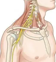
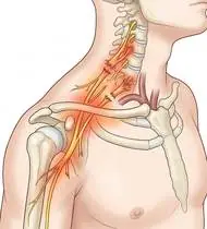
Brachial Plexus Injury (Traumatic C5-T1) — Overview
Traumatic Brachial Plexus Injury (BPI) in adults and older children is a devastating peripheral nerve lesion caused by high-energy traction compressing or widening the cervix-to-shoulder angle (motorcycle/high-speed crashes accounting for ~70%, contact sports 'stingers/burners', or shoulder dislocations/clavicle fractures). Clinically stratified by anatomical level: 1) Upper Trunk (C5–C6 ± C7 / Erb-type): paralysis of shoulder abduction/external rotation (deltoid, supraspinatus, infraspinatus) and elbow flexion/supination (biceps, brachialis, supinator) with preserved hand function; 2) Lower Trunk (C8–T1 / Klumpke-type): intrinsic hand & wrist/finger flexor paralysis ('claw hand'); 3) Pan-Plexus (C5–T1): complete flaccid 'flail arm'. Nerve recovery is governed by Seddon (Neurapraxia, Axonotmesis, Neurotmesis) / Sunderland (I–V) grading and pre-ganglionic root avulsion vs. post-ganglionic rupture!
A brachial plexus injury (shoulder & arm nerve network injury) is a stretching, tearing, or compression of the bundle of 5 major nerves (C5 to T1) that exit the neck and travel under the collarbone into the shoulder, arm, and hand. Because nerves regrow slowly from the neck downward at about 1 millimeter per day (3 cm per month), keeping every joint in your shoulder, elbow, wrist, and fingers completely supple and flexible ensures that when the nerve signal arrives, the joint is ready to move!
Primary Causes & Biomechanics
Key Points You Should Know
- •High-velocity traction driving the shoulder inferiorly while the head/neck is forced contralaterally (Upper Trunk C5–C6), or hyperabduction traction pulling the arm overhead (Lower Trunk C8–T1)
- •Pre-Ganglionic Root Avulsion (Poor Spontaneous Prognosis — Requires Nerve Transfer!) vs. Post-Ganglionic Lesion: Horner's Syndrome (ptosis, miosis, anhidrosis from T1 sympathetic chain avulsion), winged scapula (long thoracic nerve C5–C7), and elevated hemidiaphragm (phrenic nerve C3–C5) indicate proximal root avulsion!
- •Wallerian Degeneration & 1 mm/day Axonal Regeneration Law: Because axons regenerate at ~1 mm/day (1 inch/month), maintaining 100% passive joint ROM and motor endplate viability via Electrical Stimulation (EMS) while waiting for reinnervation is mandatory!
- •Why Joint Flexibility Cannot Wait for the Nerve: A nerve takes months to grow down the arm; if the shoulder, elbow, or fingers stiffen up while waiting, even a healed nerve won't be able to move a frozen joint! Do gentle assisted movements 3–4 times every single day.
- •Support the Arm Weight (Never Let It Hang!): When shoulder muscles are weak, the weight of your arm pulls the shoulder ball down out of its socket and stretches the healing nerves—always wear your supportive sling when walking.
- •Protecting Numb Skin From Burns: If parts of your arm or hand lack feeling, always look with your eyes when near stoves, hot water, or sharp edges.
Assessment Methods
1. Goniometric ROM & Postural / Kinematic Analysis
Detailed MRC (0–5) Manual Muscle Testing across all C5–T1 myotomes (Rhomboids/Serratus, Supraspinatus/Deltoid, Infraspinatus, Biceps/Brachioradialis, Triceps/Wrist Extensors, Finger Flexors, Interossei) and C5–T1 dermatomal sensory mapping.
2. Special Clinical Provocation & Neurological/Functional Tests
Advancing Tinel's Sign along the supraclavicular/infraclavicular plexus (confirming post-ganglionic axonal regeneration!), Glenohumeral Inferior Subluxation Sulcus check, and Horner's Syndrome screen.
3. Standardized Clinical Outcome Scales & Imaging Criteria
MRC Muscle Grading, DASH Score, EMG/NCV (performed at 3–4 weeks post-injury when fibrillation potentials appear), and Cervical CT Myelogram / Brachial Plexus MR Neurography (detecting pseudomeningoceles).
1. Range of Motion & Posture Check
Your physiotherapist gently evaluates how comfortably and smoothly your brachial plexus network (C5, C6, C7, C8, T1 spinal nerve roots, upper/middle/lower trunks, divisions, cords, and terminal branches) moves and checks your overall posture.
2. Muscle Strength & Functional Movement Check
Your muscle balance, walking or reaching comfort, and specific gentle movement tests are evaluated without triggering sharp pain.
3. Typical Symptoms & Daily Impact Review
Unilateral weakness or paralysis of the shoulder and elbow (Upper Plexus C5–C6), the hand and fingers (Lower Plexus C8–T1), or the entire arm (Pan-Plexus C5–T1) following trauma.
Treatment Options & Phase-Based Protocol
✨ Enriched GIF Exercise & Precaution GuidePhase 1: Acute Symptom Modulation, Tissue Protection & Early Activation
- •Phase 1 Clinical Focus & Tissue Protection: Phase 1 — Flaccid Protection, Subluxation Slings & Full Passive ROM Phase: Support the flaccid arm in a custom brachial plexus sling/hemi-sling (preventing glenohumeral subluxation & traction on regenerating nerves!), perform daily full-arc passive/wand-assisted shoulder elevation, elbow ROM, forearm supination-pronation, and Six-Pack tendon gliding & intrinsic-plus hand splinting.
- •Pain, Edema & Neuromuscular Modulation: Targeted physical modalities, relative load management, and early protected mobility of the brachial plexus network (C5, C6, C7, C8, T1 spinal nerve roots, upper/middle/lower trunks, divisions, cords, and terminal branches) without provoking mechanical or inflammatory irritation.
- •Proximal & Distal Kinetic Chain Maintenance: Maintain active mobility and circulation in adjacent joints while protecting the primary healing structures in Brachial Plexus Injury.
Phase 2: Progressive Mobility, Motor Control & Targeted Strengthening
- •Phase 2 Progressive Mobilization & Motor Control: Phase 2 — Gravity-Eliminated Reinnervation & NMES Phase: Use long-pulse Denervated Muscle Electrical Stimulation / NMES, scapular retraction, gravity-eliminated supine/table-slide elbow flexion & shoulder external rotation (Towel ER) as nascent motor units (MRC 1 -> 2) reinnervate.
- •Biomechanical Alignment & Co-Contraction: Restore balanced agonist-antagonist force couples, joint centration, and multi-planar neuromuscular control across the brachial plexus network (C5, C6, C7, C8, T1 spinal nerve roots, upper/middle/lower trunks, divisions, cords, and terminal branches).
- •Myofascial Extensibility & Endurance: Address secondary compensatory tightness and build local muscular endurance within safe, symptom-free biomechanical parameters.
Phase 3: Functional Integration, Proprioception & Long-Term Prevention
- •Phase 3 Functional Integration & Long-Term Prevention: Phase 3 — Antigravity Progressive Strengthening & Post-Nerve-Transfer Cortical Re-Education Phase: Side-lying external rotation, Full-Can elevation, thumb opposition & finger spreading, grip strengthening, and Donor-Nerve Co-Activation training (e.g., after Oberlin ulnar-to-musculocutaneous nerve transfer, squeezing the ulnar fingers while flexing the elbow!).
- •Proprioceptive Balance & Dynamic Loading: Progress to task-specific closed-chain, balance, and functional movement patterns tailored to daily life, occupational, or athletic demands.
- •Self-Management & Relapse Prophylaxis: Establish lifelong ergonomic habits, home exercise maintenance, and early warning-sign awareness to prevent recurrence.
Step 1: Calming Pain, Protecting the Area & Gentle Starter Motion
- •Why Joint Flexibility Cannot Wait for the Nerve: A nerve takes months to grow down the arm; if the shoulder, elbow, or fingers stiffen up while waiting, even a healed nerve won't be able to move a frozen joint! Do gentle assisted movements 3–4 times every single day.
- •Calm Protection & Gentle Start: During the initial stage of Shoulder & Arm Nerve Network Injury, avoid painful overloading and perform the gentle, pain-free starter movements shown below to support circulation and comfort.
- •Respecting Your Body's Signals: Every movement should stay within a comfortable, non-painful zone—never force through sharp or radiating pain.
Step 2: Restoring Flexibility & Building Supporting Muscle Strength
- •Support the Arm Weight (Never Let It Hang!): When shoulder muscles are weak, the weight of your arm pulls the shoulder ball down out of its socket and stretches the healing nerves—always wear your supportive sling when walking.
- •Step-by-Step Flexibility & Support: As acute sensitivity settles, gradually build mobility and strengthen the supporting muscles around the joint at a steady, controlled pace.
- •Posture & Breathing Coordination: Keep your posture upright and breathe smoothly (never hold your breath!) during every repetition.
Step 3: Safe Daily Habits, Stamina & Preventing Recurrence
- •Protecting Numb Skin From Burns: If parts of your arm or hand lack feeling, always look with your eyes when near stoves, hot water, or sharp edges.
- •Confident Return to Daily Life: Integrate your stronger muscles and improved balance into walking, stairs, work, and recreational activities.
- •Preventing Future Flare-Ups: Continue your core protective habits and key maintenance exercises even after symptoms resolve.
Animated (GIF) Therapeutic Exercise Guide
Animated clinical exercise protocol with biomechanical angles, target muscle groups, and dosage parameters (Click image to enlarge)
Step-by-step animated exercise demonstrations you can safely perform at home (Click image to enlarge)
 🔍 Enlarge
1. Supine Wand-Assisted (Cane) Active-Assisted Shoulder Elevation
1. Lying Cane/Stick-Assisted Two-Handed Arm Raise
In supine (scapula stabilized against the plinth and gravity eliminated in the final arc), the unaffected upper extremity guides a wand/stick to elevate the affected shoulder smoothly without deltoid-induced superior humeral shear. Specifically indicated in Brachial Plexus Injury rehabilitation to optimize brachial plexus network (C5, C6, C7, C8, T1 spinal nerve roots, upper/middle/lower trunks, divisions, cords, and terminal branches) biomechanics and neuromuscular control.
Lie on your back holding a light stick or cane with both hands. Use your healthy arm to gently guide both arms up overhead as far as comfortable, hold for 3 seconds, and slowly lower. Perform smoothly within a comfortable, pain-free range to support your Shoulder & Arm Nerve Network Injury recovery.
 🔍 Enlarge
2. Towel-Supported Glenohumeral External Rotation (Infraspinatus & Teres Minor)
2. Outward Forearm Rotation with Rolled Towel Under Elbow
External rotation performed at 90° elbow flexion with a rolled towel placed between the elbow and trunk (20°–30° scaption). Enhances infraspinatus/teres minor recruitment while preventing superior humeral head migration and deltoid shear. Specifically indicated in Brachial Plexus Injury rehabilitation to optimize brachial plexus network (C5, C6, C7, C8, T1 spinal nerve roots, upper/middle/lower trunks, divisions, cords, and terminal branches) biomechanics and neuromuscular control.
Place a small rolled towel between your elbow and your side with your elbow bent at 90 degrees. Keeping your elbow pressed gently against the towel, rotate your forearm outward, hold for 3 seconds, and return slowly. Perform smoothly within a comfortable, pain-free range to support your Shoulder & Arm Nerve Network Injury recovery.
 🔍 Enlarge
3. Scapulothoracic Retraction & Depression (Middle/Lower Trapezius & Rhomboids)
3. Shoulder Blade Squeeze (Postural Alignment)
Adduction and depression of the scapulae via the middle/lower trapezius and rhomboids while inhibiting upper trapezius overactivity. Corrects anterior scapular tilt, widens the subacromial outlet, and unloads the anterior thoracic column. Specifically indicated in Brachial Plexus Injury rehabilitation to optimize brachial plexus network (C5, C6, C7, C8, T1 spinal nerve roots, upper/middle/lower trunks, divisions, cords, and terminal branches) biomechanics and neuromuscular control.
Sit or stand tall. Keep your shoulders relaxed away from your ears, gently draw your shoulder blades together and slightly downward toward your back pockets, hold for 5 seconds, and release. Perform smoothly within a comfortable, pain-free range to support your Shoulder & Arm Nerve Network Injury recovery.
 🔍 Enlarge
4. Isolated Humeroulnar Active/Active-Assisted Flexion and Extension ROM
4. Full Elbow Bending and Straightening
Controlled full-arc elbow flexion and extension with the humerus stabilized in neutral alignment. Prevents anterior capsular contracture (extension loss) and restores biceps/triceps reciprocal excursion. Specifically indicated in Brachial Plexus Injury rehabilitation to optimize brachial plexus network (C5, C6, C7, C8, T1 spinal nerve roots, upper/middle/lower trunks, divisions, cords, and terminal branches) biomechanics and neuromuscular control.
Keep your upper arm comfortably close to your body. Slowly bend your elbow to bring your hand toward your shoulder, hold for 3 seconds, then slowly straighten your elbow as far as comfortable. Perform smoothly within a comfortable, pain-free range to support your Shoulder & Arm Nerve Network Injury recovery.
 🔍 Enlarge
5. Proximal & Distal Radioulnar Joint (PRUJ/DRUJ) Supination and Pronation
5. Elbow-at-Side Palm Up and Palm Down Rotation
Isolated forearm rotation with the elbow flexed at 90° and adducted against the trunk to eliminate compensatory glenohumeral rotation. Restores radial head spinning and distal radioulnar translation. Specifically indicated in Brachial Plexus Injury rehabilitation to optimize brachial plexus network (C5, C6, C7, C8, T1 spinal nerve roots, upper/middle/lower trunks, divisions, cords, and terminal branches) biomechanics and neuromuscular control.
Keep your elbow bent at a right angle and tucked closely against your waist. Slowly turn your palm upward toward the ceiling, hold for 3 seconds, then turn your palm downward toward the floor. Perform smoothly within a comfortable, pain-free range to support your Shoulder & Arm Nerve Network Injury recovery.
 🔍 Enlarge
6. Six-Pack Differential Flexor Tendon Gliding (FDS & FDP Excursion)
6. Step-by-Step Finger Tendon Gliding (Hook, Straight & Full Fist)
Sequential progression through Straight Hand, Tabletop (intrinsic-plus), Hook Fist (maximum FDS/FDP differential glide), Full Fist, and Straight Fist positions. Prevents peritendinous adhesions within the carpal tunnel and fibro-osseous digital sheaths. Specifically indicated in Brachial Plexus Injury rehabilitation to optimize brachial plexus network (C5, C6, C7, C8, T1 spinal nerve roots, upper/middle/lower trunks, divisions, cords, and terminal branches) biomechanics and neuromuscular control.
Start with your fingers straight, then bend at the knuckles (tabletop), curl just the fingertips (hook fist), make a gentle full fist, and open wide again—holding each position for 2–3 seconds. Perform smoothly within a comfortable, pain-free range to support your Shoulder & Arm Nerve Network Injury recovery.
Movements to Avoid & Strict Precautions
1. Letting a Paralyzed/Weak Arm Dangle Unsupported While Standing or Walking (Re-Stretches the Healing Brachial Plexus!)
1. Letting your weak arm hang unsupported at your side while walking (pulls on the shoulder joint and healing nerves!)
2. Forcefully Pulling the Neck Away From the Injured Shoulder (Traction Stress on C5–T1 Roots!)
2. Forcefully tilting your head away from the injured shoulder while pulling the arm down
3. Waiting More Than 3–6 Months With Zero Muscle Recovery Without Seeing a Brachial Plexus Microsurgeon
3. Waiting over a year without specialist nerve evaluation when a muscle shows zero recovery at 3–6 months
4. Touching Hot Pans, Radiators, or Hot Bathwater With an Insensate Hand
4. Handling hot objects or bathwater with a hand that lacks temperature sensation
Brachial Plexus Injury (Traumatic C5-T1) — Video Guide
Click directly on any video to open and watch in a window, or review details via 📋 Summary & Options.
Condition Overview Video (1 Video)
Summary of pathophysiology, biomechanics, and clinical presentation
What the condition is, why it develops, and its basic mechanism
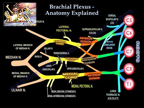
What Is a Brachial Plexus Injury? C5–T1 Nerve Network Anatomy and Symptoms
Traumatic Brachial Plexus Injury (BPI) in adults and older children is a devastating peripheral nerve lesion caused by high-energy traction compressing or widening the cervix-to-shoulder angle (motorcycle/high-speed crashes accounting for ~70%, contact sports 'stingers/burners', or shoulder dislocations/clavicle fractures). Clinically stratified by anatomical level: 1) Upper Trunk (C5–C6 ± C7 / Erb-type): paralysis of shoulder abduction/external rotation (deltoid, supraspinatus, infraspinatus) and elbow flexion/supination (biceps, brachialis, supinator) with preserved hand function; 2) Lower Trunk (C8–T1 / Klumpke-type): intrinsic hand & wrist/finger flexor paralysis ('claw hand'); 3) Pan-Plexus (C5–T1): complete flaccid 'flail arm'. Nerve recovery is governed by Seddon (Neurapraxia, Axonotmesis, Neurotmesis) / Sunderland (I–V) grading and pre-ganglionic root avulsion vs. post-ganglionic rupture!
A brachial plexus injury (shoulder & arm nerve network injury) is a stretching, tearing, or compression of the bundle of 5 major nerves (C5 to T1) that exit the neck and travel under the collarbone into the shoulder, arm, and hand. Because nerves regrow slowly from the neck downward at about 1 millimeter per day (3 cm per month), keeping every joint in your shoulder, elbow, wrist, and fingers completely supple and flexible ensures that when the nerve signal arrives, the joint is ready to move!
Treatment and Exercise Videos
Phase-based physiotherapy protocols, mobilization, and therapeutic exercises
Step-by-step recovery and safe home exercise applications

Upper Extremity Range of Motion Exercises in Brachial Plexus Injuries
Phase 1: Acute Symptom Modulation, Tissue Protection & Early Activation: Phase 1 Clinical Focus & Tissue Protection: Phase 1 — Flaccid Protection, Subluxation Slings & Full Passive ROM Phase: Support the flaccid arm in a custom brachial plexus sling/hemi-sling (preventing glenohumeral subluxation & traction on regenerating nerves!), perform daily full-arc passive/wand-assisted shoulder elevation, elbow ROM, forearm supination-pronation, and Six-Pack tendon gliding & intrinsic-plus hand splinting.
Step 1: Calming Pain, Protecting the Area & Gentle Starter Motion: Why Joint Flexibility Cannot Wait for the Nerve: A nerve takes months to grow down the arm; if the shoulder, elbow, or fingers stiffen up while waiting, even a healed nerve won't be able to move a frozen joint! Do gentle assisted movements 3–4 times every single day.

Pediatric Brachial Plexus (Erb-Duchenne & Klumpke) Rehabilitation Exercises
Phase 2: Progressive Mobility, Motor Control & Targeted Strengthening: Phase 2 Progressive Mobilization & Motor Control: Phase 2 — Gravity-Eliminated Reinnervation & NMES Phase: Use long-pulse Denervated Muscle Electrical Stimulation / NMES, scapular retraction, gravity-eliminated supine/table-slide elbow flexion & shoulder external rotation (Towel ER) as nascent motor units (MRC 1 -> 2) reinnervate.
Step 2: Restoring Flexibility & Building Supporting Muscle Strength: Support the Arm Weight (Never Let It Hang!): When shoulder muscles are weak, the weight of your arm pulls the shoulder ball down out of its socket and stretches the healing nerves—always wear your supportive sling when walking.
Clinical Assessment, Goniometry & Special Test Videos
Displayed only in Healthcare Professional mode (hidden in Patient mode)
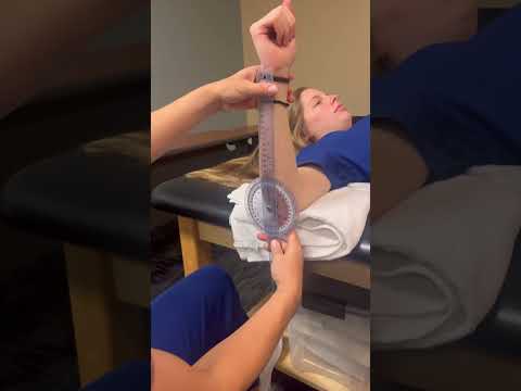
Shoulder Joint Goniometric Measurement (Flexion, Abduction, Internal & External Rotation)
Goniometric ROM & Postural / Kinematic Analysis: Detailed MRC (0–5) Manual Muscle Testing across all C5–T1 myotomes (Rhomboids/Serratus, Supraspinatus/Deltoid, Infraspinatus, Biceps/Brachioradialis, Triceps/Wrist Extensors, Finger Flexors, Interossei) and C5–T1 dermatomal sensory mapping.

Manual Muscle Testing (MRC 0–5 Grading) — Clinical Motor Strength Examination
Special Clinical Provocation & Neurological/Functional Tests: Advancing Tinel's Sign along the supraclavicular/infraclavicular plexus (confirming post-ganglionic axonal regeneration!), Glenohumeral Inferior Subluxation Sulcus check, and Horner's Syndrome screen.

Systematic Neurological Examination: Reflex, Sensory, Tone, and Motor Assessment
Standardized Clinical Outcome Scales & Imaging Criteria: MRC Muscle Grading, DASH Score, EMG/NCV (performed at 3–4 weeks post-injury when fibrillation potentials appear), and Cervical CT Myelogram / Brachial Plexus MR Neurography (detecting pseudomeningoceles).