Pneumonia (Alveolar Consolidation)Pneumonia / Lung Infection (Pneumonia)
Pneumonia (Alveolar Consolidation) — Overview
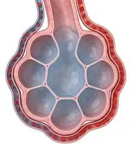
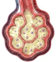
Pneumonia is an acute lower respiratory tract infection of the lung parenchyma (alveoli and terminal bronchioles) caused by bacterial (*Streptococcus pneumoniae*), viral, or aspiration pathogens. Inflammatory exudate (fibrin, neutrophils, edema fluid, and cellular debris) fills the alveolar spaces (Alveolar Consolidation), resulting in loss of functional gas-exchange surface area, reduced lung compliance, localized basal/lobar atelectasis, and intrapulmonary shunting (V/Q mismatch -> hypoxemia). Crucially, physiotherapy differs by stage: during the early dry/febrile stage (consolidation without loose secretions), chest percussion is ineffective and exhausting—focus is on upright positioning ('Good Lung Down' in unilateral pneumonia to maximize V/Q matching!), localized Thoracic Expansion Exercises (TEE), Incentive Spirometry (Triflo), and early mobilization; during the resolution/productive stage, ACBT and Huffing clear resolving exudate!
Pneumonia (lung infection) occurs when an infection causes the tiny air sacs (alveoli) in a section (lobe) of your lung to fill with fluid and mucus instead of fresh air—making breathing feel shallow and tiring. Alongside your prescribed antibiotics/medications, sitting upright out of bed, doing hourly slow deep-inhalation breaths (Incentive Spirometer / Triflo), and gentle 'huffing' re-inflates the collapsed air sacs and speeds up recovery!
Primary Causes & Biomechanics
Key Points You Should Know
- •Alveolar filling with inflammatory exudate reducing pulmonary compliance and blocking oxygen diffusion across the alveolar-capillary membrane
- •Ventilation-Perfusion (V/Q) Gravity Rule in Unilateral Lobar Pneumonia: In adults, gravity directs the greatest blood flow (perfusion) to the dependent (bottom) lung! Therefore, positioning the patient in side-lying with the HEALTHY LUNG DOWN ('Good Lung Down') matches maximum perfusion with healthy ventilation (boosting SpO2 immediately!) while placing the consolidated lobe UP to promote bronchial drainage and expansion!
- •Prolonged supine bed rest during hospitalization causing posterior basal atelectasis, DVT, and rapid 2–5%/day quadriceps sarcopenia
- •How to Use Your 3-Ball Breathing Device (Triflo / Incentive Spirometer) Correctly: The #1 mistake people make with a 3-ball Triflo device is trying to *blow into it* or sucking in as fast as a vacuum! You must BREATHE IN (inhale) slowly and steadily (like sipping a thick milkshake through a straw) so the balls float gently, then HOLD that breath inside for 3 to 5 seconds before exhaling! That 3-second hold pushes air through tiny side-doors (collateral channels) to pop open stuck air sacs!
- •Sitting Upright in a Chair Beats Lying Flat in Bed: When you lie flat on your back, the weight of your belly presses against the bottom of your lungs. Sitting upright in an armchair (or lying on your healthy side) immediately opens the lung bases and raises your oxygen level!
- •Splint Your Ribs With a Pillow When Coughing: If deep breathing or coughing causes a sharp stitch in your side (pleuritic pain), hug a small firm pillow against that side of your ribs while doing a gentle 'Huff'.
Assessment Methods
1. Goniometric ROM & Postural / Kinematic Analysis
Auscultation (localized bronchial breath sounds, fine end-inspiratory crackles/rales, and increased tactile vocal fremitus/dull percussion over the consolidated lobe), Respiratory Rate (normal: 12–20/min), and Pulse Oximetry (SpO2).
2. Special Clinical Provocation & Neurological/Functional Tests
CURB-65 Severity Score (Confusion, Urea, Respiratory rate ≥30, BP <90/60, Age ≥65), Segmental Chest Expansion Palpation (manual tactile cueing over the affected basal/middle/upper lobe), and 6-Minute Walk / Sit-to-Stand tolerance.
3. Standardized Clinical Outcome Scales & Imaging Criteria
SpO2 (%), Respiratory Rate, Borg Dyspnea Scale, Chest X-Ray lobar resolution, and 30-Second Sit-to-Stand.
1. Range of Motion & Posture Check
Your physiotherapist gently evaluates how comfortably and smoothly your pulmonary alveoli and respiratory bronchioles (inflammatory exudate, consolidation, and ventilation/perfusion [V/Q] mismatch) moves and checks your overall posture.
2. Muscle Strength & Functional Movement Check
Your muscle balance, walking or reaching comfort, and specific gentle movement tests are evaluated without triggering sharp pain.
3. Typical Symptoms & Daily Impact Review
Fever, Chills, Productive Cough (rusty, yellow, or green phlegm), and shortness of breath with rapid shallow breathing.
Treatment Options & Phase-Based Protocol
✨ Enriched GIF Exercise & Precaution GuidePhase 1: Acute Symptom Modulation, Tissue Protection & Early Activation
- •Phase 1 Clinical Focus & Tissue Protection: Phase 1 — Acute / Early Consolidation Phase (Positioning, Oxygenation & Atelectasis Prevention): Elevate head of bed ≥30–45° or sit upright in a chair (never leave flat on the back!), use 'Good Lung Down' side-lying positioning, perform hourly Incentive Spirometry (Triflo: slow deep *INHALATION*—never blowing out!—with a 3–5s end-inspiratory hold to open collateral Pores of Kohn!), localized tactile Thoracic Expansion Exercises (placing hands over the affected ribcage), and hourly ankle pumps.
- •Pain, Edema & Neuromuscular Modulation: Targeted physical modalities, relative load management, and early protected mobility of the pulmonary alveoli and respiratory bronchioles (inflammatory exudate, consolidation, and ventilation/perfusion [V/Q] mismatch) without provoking mechanical or inflammatory irritation.
- •Proximal & Distal Kinetic Chain Maintenance: Maintain active mobility and circulation in adjacent joints while protecting the primary healing structures in Pneumonia.
Phase 2: Progressive Mobility, Motor Control & Targeted Strengthening
- •Phase 2 Progressive Mobilization & Motor Control: Phase 2 — Productive Resolution Phase (Airway Clearance & Bedside Mobilization): Once alveolar exudate liquefies (wet crackles/productive cough), perform Active Cycle of Breathing Techniques (ACBT) + open-glottis Huffing (splinting the ribcage with a pillow if pleuritic pain is present!), unloaded supine heel slides, and supported chair sit-to-stand & hallway walking.
- •Biomechanical Alignment & Co-Contraction: Restore balanced agonist-antagonist force couples, joint centration, and multi-planar neuromuscular control across the pulmonary alveoli and respiratory bronchioles (inflammatory exudate, consolidation, and ventilation/perfusion [V/Q] mismatch).
- •Myofascial Extensibility & Endurance: Address secondary compensatory tightness and build local muscular endurance within safe, symptom-free biomechanical parameters.
Phase 3: Functional Integration, Proprioception & Long-Term Prevention
- •Phase 3 Functional Integration & Long-Term Prevention: Phase 3 — Post-Discharge Lung Capacity & Deconditioning Recovery Phase (Weeks 2–6): Bilateral arm elevation with deep costal expansion, scapular retraction, pelvic bridging & wall mini-squats, and progressive daily walking (noting that post-pneumonia fatigue normally takes 4–6 weeks to fully clear!).
- •Proprioceptive Balance & Dynamic Loading: Progress to task-specific closed-chain, balance, and functional movement patterns tailored to daily life, occupational, or athletic demands.
- •Self-Management & Relapse Prophylaxis: Establish lifelong ergonomic habits, home exercise maintenance, and early warning-sign awareness to prevent recurrence.
Step 1: Calming Pain, Protecting the Area & Gentle Starter Motion
- •How to Use Your 3-Ball Breathing Device (Triflo / Incentive Spirometer) Correctly: The #1 mistake people make with a 3-ball Triflo device is trying to *blow into it* or sucking in as fast as a vacuum! You must BREATHE IN (inhale) slowly and steadily (like sipping a thick milkshake through a straw) so the balls float gently, then HOLD that breath inside for 3 to 5 seconds before exhaling! That 3-second hold pushes air through tiny side-doors (collateral channels) to pop open stuck air sacs!
- •Calm Protection & Gentle Start: During the initial stage of Pneumonia / Lung Infection, avoid painful overloading and perform the gentle, pain-free starter movements shown below to support circulation and comfort.
- •Respecting Your Body's Signals: Every movement should stay within a comfortable, non-painful zone—never force through sharp or radiating pain.
Step 2: Restoring Flexibility & Building Supporting Muscle Strength
- •Sitting Upright in a Chair Beats Lying Flat in Bed: When you lie flat on your back, the weight of your belly presses against the bottom of your lungs. Sitting upright in an armchair (or lying on your healthy side) immediately opens the lung bases and raises your oxygen level!
- •Step-by-Step Flexibility & Support: As acute sensitivity settles, gradually build mobility and strengthen the supporting muscles around the joint at a steady, controlled pace.
- •Posture & Breathing Coordination: Keep your posture upright and breathe smoothly (never hold your breath!) during every repetition.
Step 3: Safe Daily Habits, Stamina & Preventing Recurrence
- •Splint Your Ribs With a Pillow When Coughing: If deep breathing or coughing causes a sharp stitch in your side (pleuritic pain), hug a small firm pillow against that side of your ribs while doing a gentle 'Huff'.
- •Confident Return to Daily Life: Integrate your stronger muscles and improved balance into walking, stairs, work, and recreational activities.
- •Preventing Future Flare-Ups: Continue your core protective habits and key maintenance exercises even after symptoms resolve.
Animated (GIF) Therapeutic Exercise Guide
Animated clinical exercise protocol with biomechanical angles, target muscle groups, and dosage parameters (Click image to enlarge)
Step-by-step animated exercise demonstrations you can safely perform at home (Click image to enlarge)
 🔍 Enlarge
1. Diaphragmatic & Pursed-Lip Breathing (PLB — Autonomic & Ventilatory Control)
1. Calm Belly Breathing with Pursed-Lip Exhale
Slow nasal inhalation expanding the lower ribcage and abdomen (while relaxing accessory neck/shoulder muscles), followed by prolonged expiration through pursed lips at a 1:2 ratio. Prevents dynamic airway collapse, improves basal ventilation, and reduces sympathetic tone. Specifically indicated in Pneumonia rehabilitation to optimize pulmonary alveoli and respiratory bronchioles (inflammatory exudate, consolidation, and ventilation/perfusion [V/Q] mismatch) biomechanics and neuromuscular control.
Sit comfortably and place one hand on your chest and the other on your belly. Breathe in slowly through your nose for 2–3 seconds so your belly gently rises, then breathe out slowly for 4–6 seconds through pursed lips as if flickering a candle. Perform smoothly within a comfortable, pain-free range to support your Pneumonia / Lung Infection recovery.
 🔍 Enlarge
2. Rhythmic Active Ankle Dorsiflexion & Plantarflexion Pumping (Venous/Edema Pump)
2. Ankle Toe-Pulls and Pointing (Circulation & Swelling Pump)
Rhythmic alternating contraction of the tibialis anterior and gastrocnemius-soleus complex. Activates the deep venous calf pump to prevent deep vein thrombosis (DVT), clears distal edema, and maintains talocrural mobility. Specifically indicated in Pneumonia rehabilitation to optimize pulmonary alveoli and respiratory bronchioles (inflammatory exudate, consolidation, and ventilation/perfusion [V/Q] mismatch) biomechanics and neuromuscular control.
While lying down or sitting with your legs supported, pull your toes and feet firmly upward toward your shins, hold for 3 seconds, then point them downward like pressing a gas pedal. Perform smoothly within a comfortable, pain-free range to support your Pneumonia / Lung Infection recovery.
 🔍 Enlarge
3. Thoracic Expansion Exercise (TEE) with Bilateral Arm Elevation & 3s Inspiratory Hold
3. Chest-Opening Deep Breath with Arm Raise
Synchronizing deep nasal inspiration with bilateral shoulder flexion/abduction and a 3-second end-inspiratory hold before passive/pursed-lip expiration. Opens collateral ventilation channels to prevent basal atelectasis and maintains costovertebral compliance. Specifically indicated in Pneumonia rehabilitation to optimize pulmonary alveoli and respiratory bronchioles (inflammatory exudate, consolidation, and ventilation/perfusion [V/Q] mismatch) biomechanics and neuromuscular control.
While sitting upright, breathe in deeply through your nose while opening your arms upward and outward to expand your ribcage. Hold the breath inside for 3 seconds, then exhale slowly while lowering your arms. Perform smoothly within a comfortable, pain-free range to support your Pneumonia / Lung Infection recovery.
 🔍 Enlarge
4. Functional Closed-Kinetic-Chain Chair Transfer (Sit-to-Stand Biomechanics)
4. Controlled Chair Sit-to-Stand Transfer
Controlled transition from sitting to standing and eccentric lowering back to the chair with symmetrical weight distribution, anterior trunk inclination ('nose over toes'), and neutral frontal knee alignment. Specifically indicated in Pneumonia rehabilitation to optimize pulmonary alveoli and respiratory bronchioles (inflammatory exudate, consolidation, and ventilation/perfusion [V/Q] mismatch) biomechanics and neuromuscular control.
Sit near the front of a sturdy chair with your feet hip-width apart. Lean slightly forward and push evenly through both heels to stand up tall, then slowly lower yourself back down over 3 seconds. Perform smoothly within a comfortable, pain-free range to support your Pneumonia / Lung Infection recovery.
 🔍 Enlarge
5. Supine Active-Assisted Heel Slide & Synovial Diffusion Mobilization
5. Supine Heel Slide (Gentle Knee & Hip Bending)
Controlled flexion and extension of the lower extremity by sliding the heel along the support surface without axial weight-bearing. Stimulates synovial fluid diffusion across avascular cartilage and prevents capsular adhesions. Specifically indicated in Pneumonia rehabilitation to optimize pulmonary alveoli and respiratory bronchioles (inflammatory exudate, consolidation, and ventilation/perfusion [V/Q] mismatch) biomechanics and neuromuscular control.
While lying on your back, slowly slide your heel along the bed sheet toward your hip within a comfortable, pain-free angle, hold for 3 seconds, and gently slide it back out straight. Perform smoothly within a comfortable, pain-free range to support your Pneumonia / Lung Infection recovery.
 🔍 Enlarge
6. Supine Wand-Assisted (Cane) Active-Assisted Shoulder Elevation
6. Lying Cane/Stick-Assisted Two-Handed Arm Raise
In supine (scapula stabilized against the plinth and gravity eliminated in the final arc), the unaffected upper extremity guides a wand/stick to elevate the affected shoulder smoothly without deltoid-induced superior humeral shear. Specifically indicated in Pneumonia rehabilitation to optimize pulmonary alveoli and respiratory bronchioles (inflammatory exudate, consolidation, and ventilation/perfusion [V/Q] mismatch) biomechanics and neuromuscular control.
Lie on your back holding a light stick or cane with both hands. Use your healthy arm to gently guide both arms up overhead as far as comfortable, hold for 3 seconds, and slowly lower. Perform smoothly within a comfortable, pain-free range to support your Pneumonia / Lung Infection recovery.
Movements to Avoid & Strict Precautions
1. Exercising While Running a High Fever (>38°C / 100.4°F — Risk of Viral Myocarditis & Dehydration!)
1. Doing strenuous exercise while you still have an active fever (focus only on upright sitting, gentle deep breaths, and ankle pumps until fever clears!)
2. Blowing Out Into an Incentive Spirometer (Triflo) or Sucking In Violently Fast
2. Blowing into your Triflo device instead of inhaling slowly and holding for 3–5 seconds
3. Lying Flat on Your Back All Day in Bed (Collapses the Lower Lung Lobes!)
3. Staying flat on your back in bed all day (sit upright in a chair several times a day!)
4. Rushing Back to Full-Intensity Sports in Week 1 Post-Pneumonia (Wait for Full 4-Week Gradual Recovery)
4. Jumping straight into intense workouts the day after finishing antibiotics without gradual walking progression
Pneumonia (Alveolar Consolidation) — Video Guide
Click directly on any video to open and watch in a window, or review details via 📋 Summary & Options.
Condition Overview Video (1 Video)
Summary of pathophysiology, biomechanics, and clinical presentation
What the condition is, why it develops, and its basic mechanism

What Is Pneumonia? Preserving Lung Capacity During Recovery
Pneumonia is an acute lower respiratory tract infection of the lung parenchyma (alveoli and terminal bronchioles) caused by bacterial (*Streptococcus pneumoniae*), viral, or aspiration pathogens. Inflammatory exudate (fibrin, neutrophils, edema fluid, and cellular debris) fills the alveolar spaces (Alveolar Consolidation), resulting in loss of functional gas-exchange surface area, reduced lung compliance, localized basal/lobar atelectasis, and intrapulmonary shunting (V/Q mismatch -> hypoxemia). Crucially, physiotherapy differs by stage: during the early dry/febrile stage (consolidation without loose secretions), chest percussion is ineffective and exhausting—focus is on upright positioning ('Good Lung Down' in unilateral pneumonia to maximize V/Q matching!), localized Thoracic Expansion Exercises (TEE), Incentive Spirometry (Triflo), and early mobilization; during the resolution/productive stage, ACBT and Huffing clear resolving exudate!
Pneumonia (lung infection) occurs when an infection causes the tiny air sacs (alveoli) in a section (lobe) of your lung to fill with fluid and mucus instead of fresh air—making breathing feel shallow and tiring. Alongside your prescribed antibiotics/medications, sitting upright out of bed, doing hourly slow deep-inhalation breaths (Incentive Spirometer / Triflo), and gentle 'huffing' re-inflates the collapsed air sacs and speeds up recovery!
Treatment and Exercise Videos
Phase-based physiotherapy protocols, mobilization, and therapeutic exercises
Step-by-step recovery and safe home exercise applications

Lung Expansion and Atelectasis Prevention Exercises with an Incentive Spirometer (Triflo)
Phase 1: Acute Symptom Modulation, Tissue Protection & Early Activation: Phase 1 Clinical Focus & Tissue Protection: Phase 1 — Acute / Early Consolidation Phase (Positioning, Oxygenation & Atelectasis Prevention): Elevate head of bed ≥30–45° or sit upright in a chair (never leave flat on the back!), use 'Good Lung Down' side-lying positioning, perform hourly Incentive Spirometry (Triflo: slow deep *INHALATION*—never blowing out!—with a 3–5s end-inspiratory hold to open collateral Pores of Kohn!), localized tactile Thoracic Expansion Exercises (placing hands over the affected ribcage), and hourly ankle pumps.
Step 1: Calming Pain, Protecting the Area & Gentle Starter Motion: How to Use Your 3-Ball Breathing Device (Triflo / Incentive Spirometer) Correctly: The #1 mistake people make with a 3-ball Triflo device is trying to *blow into it* or sucking in as fast as a vacuum! You must BREATHE IN (inhale) slowly and steadily (like sipping a thick milkshake through a straw) so the balls float gently, then HOLD that breath inside for 3 to 5 seconds before exhaling! That 3-second hold pushes air through tiny side-doors (collateral channels) to pop open stuck air sacs!

Proper Incentive Spirometer (Triflo) Technique in Respiratory Physiotherapy
Phase 2: Progressive Mobility, Motor Control & Targeted Strengthening: Phase 2 Progressive Mobilization & Motor Control: Phase 2 — Productive Resolution Phase (Airway Clearance & Bedside Mobilization): Once alveolar exudate liquefies (wet crackles/productive cough), perform Active Cycle of Breathing Techniques (ACBT) + open-glottis Huffing (splinting the ribcage with a pillow if pleuritic pain is present!), unloaded supine heel slides, and supported chair sit-to-stand & hallway walking.
Step 2: Restoring Flexibility & Building Supporting Muscle Strength: Sitting Upright in a Chair Beats Lying Flat in Bed: When you lie flat on your back, the weight of your belly presses against the bottom of your lungs. Sitting upright in an armchair (or lying on your healthy side) immediately opens the lung bases and raises your oxygen level!
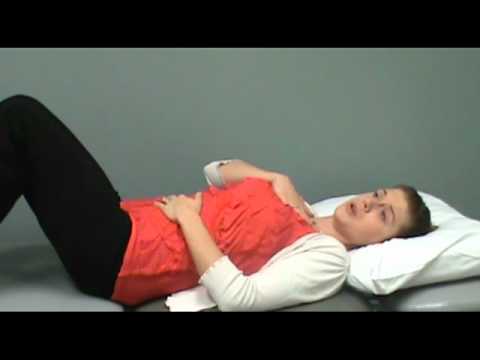
Diaphragmatic Breathing (Abdominal Breathing) Instruction and Clinical Application
Phase 3: Functional Integration, Proprioception & Long-Term Prevention: Phase 3 Functional Integration & Long-Term Prevention: Phase 3 — Post-Discharge Lung Capacity & Deconditioning Recovery Phase (Weeks 2–6): Bilateral arm elevation with deep costal expansion, scapular retraction, pelvic bridging & wall mini-squats, and progressive daily walking (noting that post-pneumonia fatigue normally takes 4–6 weeks to fully clear!).
Step 3: Safe Daily Habits, Stamina & Preventing Recurrence: Splint Your Ribs With a Pillow When Coughing: If deep breathing or coughing causes a sharp stitch in your side (pleuritic pain), hug a small firm pillow against that side of your ribs while doing a gentle 'Huff'.
Clinical Assessment, Goniometry & Special Test Videos
Displayed only in Healthcare Professional mode (hidden in Patient mode)

Systematic Respiratory Physical Examination (Inspection, Palpation, Percussion, Auscultation)
Goniometric ROM & Postural / Kinematic Analysis: Auscultation (localized bronchial breath sounds, fine end-inspiratory crackles/rales, and increased tactile vocal fremitus/dull percussion over the consolidated lobe), Respiratory Rate (normal: 12–20/min), and Pulse Oximetry (SpO2).

Why and How Is the 6-Minute Walk Test (6MWT) Performed? Functional Capacity Assessment
Special Clinical Provocation & Neurological/Functional Tests: CURB-65 Severity Score (Confusion, Urea, Respiratory rate ≥30, BP <90/60, Age ≥65), Segmental Chest Expansion Palpation (manual tactile cueing over the affected basal/middle/upper lobe), and 6-Minute Walk / Sit-to-Stand tolerance.