Interstitial Lung Diseases (ILD / Pulmonary Fibrosis)Lung Stiffening / Pulmonary Fibrosis (Interstitial Lung Diseases)
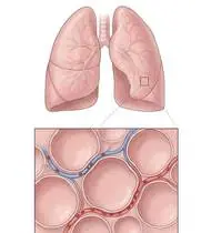
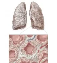
Interstitial Lung Diseases (ILD / Pulmonary Fibrosis) — Overview
Interstitial Lung Diseases (ILD — including Idiopathic Pulmonary Fibrosis [IPF], connective-tissue-associated ILD, hypersensitivity pneumonitis, and sarcoidosis) are a group of chronic parenchymal lung disorders characterized by inflammation and progressive collagen scarring (fibrosis) of the alveolar-capillary interstitium. Unlike obstructive COPD (where air is trapped and lungs are hyperinflated), ILD produces a RESTRICTIVE Ventilatory Defect (reduced TLC, FVC, and RV with a normal or elevated FEV1/FVC ratio >0.80!) and a thickened alveolar-capillary diffusion barrier (severely reduced DLCO). Consequently, the lungs become stiff and small ('like trying to inflate a thick rubber hot-water bottle instead of a balloon'), causing a rapid, shallow breathing pattern and profound, rapid Exercise-Induced Arterial Desaturation (SpO2 dropping precipitously during walking)!
Interstitial Lung Disease (ILD / pulmonary fibrosis / lung stiffening) is a condition in which the delicate lace-like tissue between the tiny air sacs in the lungs becomes thicker and stiffer (fibrosis). This makes the lungs feel less stretchy and slows down how fast oxygen crosses into the bloodstream when you walk. Pulmonary rehabilitation is proven to improve walking distance, muscle strength, and quality of life in ILD—as long as you follow one golden rule: always watch your finger pulse oximeter and pace your steps so your oxygen stays in the safe zone (≥88–90%)!
Primary Causes & Biomechanics
Key Points You Should Know
- •Fibroblastic proliferation and extracellular matrix deposition thickening the alveolar-capillary membrane and obliterating alveolar units
- •Restrictive Mechanics & High Work of Breathing: Reduced lung compliance requires high negative pleural pressure for every breath, forcing a rapid, shallow breathing frequency
- •Diffusion Limitation & Exertional Desaturation: At rest, red blood cells spend ~0.75 seconds in pulmonary capillaries (enough time for O2 to cross the thickened membrane); during exercise, cardiac output speeds up capillary transit to ~0.25 seconds—causing dramatic, rapid drops in SpO2!
- •Rule #1 — Your Pulse Oximeter Is Your Speedometer: In pulmonary fibrosis, your oxygen level can look normal while sitting still (95%), then drop quickly as soon as you walk down the hall because blood moves past the air sacs faster! Always keep a finger pulse oximeter handy during exercise, walk at a calm pace with 2-minute rest breaks, and use your prescribed portable oxygen during walking/exercise so your SpO2 stays 88% or higher!
- •How to Calm the Dry, Ticklish Cough: Taking a sudden, huge deep breath stretches the stiff lung tissue and triggers a dry coughing fit! Instead, take moderate, smooth nasal breaths, sip room-temperature water, or swallow/breathe gently through pursed lips when you feel a tickle.
- •Keep Your Ribcage Flexible: Even when the lung tissue inside is stiff, keeping your ribcage and shoulder muscles flexible makes every breath take less effort.
Assessment Methods
1. Goniometric ROM & Postural / Kinematic Analysis
Pulmonary Function Testing: Restrictive pattern (FVC <80% predicted, TLC <80%, FEV1/FVC ≥0.75–0.80) and Diffusing Capacity of the Lung for Carbon Monoxide (DLCO % predicted).
2. Special Clinical Provocation & Neurological/Functional Tests
6-Minute Walk Test (6MWT) with Continuous Pulse Oximetry (measuring the nadir SpO2 and exact oxygen flow rate in L/min needed to keep SpO2 ≥88–90% during ambulation!), mMRC / Borg Dyspnea Scale, and Chest Wall / Ribcage Compliance.
3. Standardized Clinical Outcome Scales & Imaging Criteria
FVC (% predicted), DLCO (% predicted), 6MWT Distance & Nadir SpO2 (%), and King's Brief Interstitial Lung Disease (KBILD) questionnaire.
1. Range of Motion & Posture Check
Your physiotherapist gently evaluates how comfortably and smoothly your alveolar-capillary interstitium (collagen fibrosis, thickened diffusion membrane, and restrictive lung compliance) moves and checks your overall posture.
2. Muscle Strength & Functional Movement Check
Your muscle balance, walking or reaching comfort, and specific gentle movement tests are evaluated without triggering sharp pain.
3. Typical Symptoms & Daily Impact Review
Insidious Progressive Exertional Dyspnea (Shortness of Breath on Activity) and persistent dry, non-productive hacking cough.
Treatment Options & Phase-Based Protocol
✨ Enriched GIF Exercise & Precaution GuidePhase 1: Acute Symptom Modulation, Tissue Protection & Early Activation
- •Phase 1 Clinical Focus & Tissue Protection: Phase 1 — Oxygen-Titrated Paced Breathing, Energy Conservation & Cough Suppression Phase: CONTINUOUS PULSE OXIMETRY MONITORING (Maintain SpO2 ≥88–90% at all times using titrated ambulatory supplemental oxygen if prescribed!); perform slow, paced diaphragmatic & pursed-lip breathing (avoiding forced maximal inspirations that trigger the stretch-receptor dry hacking cough reflex!), and Forward-Lean recovery postures.
- •Pain, Edema & Neuromuscular Modulation: Targeted physical modalities, relative load management, and early protected mobility of the alveolar-capillary interstitium (collagen fibrosis, thickened diffusion membrane, and restrictive lung compliance) without provoking mechanical or inflammatory irritation.
- •Proximal & Distal Kinetic Chain Maintenance: Maintain active mobility and circulation in adjacent joints while protecting the primary healing structures in Interstitial Lung Diseases.
Phase 2: Progressive Mobility, Motor Control & Targeted Strengthening
- •Phase 2 Progressive Mobilization & Motor Control: Phase 2 — Ribcage / Thoracic Compliance & Postural Expansion Phase: Because the underlying lung parenchyma is stiff, keeping the overlying thoracic cage, intercostals, and shoulder girdle as supple as possible reduces the work of breathing! Perform gentle Thoracic Expansion Exercises with arm elevation, scapular retraction, and neck/pectoral myofascial stretching.
- •Biomechanical Alignment & Co-Contraction: Restore balanced agonist-antagonist force couples, joint centration, and multi-planar neuromuscular control across the alveolar-capillary interstitium (collagen fibrosis, thickened diffusion membrane, and restrictive lung compliance).
- •Myofascial Extensibility & Endurance: Address secondary compensatory tightness and build local muscular endurance within safe, symptom-free biomechanical parameters.
Phase 3: Functional Integration, Proprioception & Long-Term Prevention
- •Phase 3 Functional Integration & Long-Term Prevention: Phase 3 — Interval-Paced Lower-Limb Strengthening & Walking Phase: Interval training (e.g., 30–60s exercise / 60s seated recovery with oxygen check!), seated terminal knee extension, chair sit-to-stand (exhaling on standing!), side-lying/standing hip abduction, and Energy Conservation in ADLs (sitting on a shower chair, dressing seated, placing everyday items at waist height).
- •Proprioceptive Balance & Dynamic Loading: Progress to task-specific closed-chain, balance, and functional movement patterns tailored to daily life, occupational, or athletic demands.
- •Self-Management & Relapse Prophylaxis: Establish lifelong ergonomic habits, home exercise maintenance, and early warning-sign awareness to prevent recurrence.
Step 1: Calming Pain, Protecting the Area & Gentle Starter Motion
- •Rule #1 — Your Pulse Oximeter Is Your Speedometer: In pulmonary fibrosis, your oxygen level can look normal while sitting still (95%), then drop quickly as soon as you walk down the hall because blood moves past the air sacs faster! Always keep a finger pulse oximeter handy during exercise, walk at a calm pace with 2-minute rest breaks, and use your prescribed portable oxygen during walking/exercise so your SpO2 stays 88% or higher!
- •Calm Protection & Gentle Start: During the initial stage of Lung Stiffening / Pulmonary Fibrosis, avoid painful overloading and perform the gentle, pain-free starter movements shown below to support circulation and comfort.
- •Respecting Your Body's Signals: Every movement should stay within a comfortable, non-painful zone—never force through sharp or radiating pain.
Step 2: Restoring Flexibility & Building Supporting Muscle Strength
- •How to Calm the Dry, Ticklish Cough: Taking a sudden, huge deep breath stretches the stiff lung tissue and triggers a dry coughing fit! Instead, take moderate, smooth nasal breaths, sip room-temperature water, or swallow/breathe gently through pursed lips when you feel a tickle.
- •Step-by-Step Flexibility & Support: As acute sensitivity settles, gradually build mobility and strengthen the supporting muscles around the joint at a steady, controlled pace.
- •Posture & Breathing Coordination: Keep your posture upright and breathe smoothly (never hold your breath!) during every repetition.
Step 3: Safe Daily Habits, Stamina & Preventing Recurrence
- •Keep Your Ribcage Flexible: Even when the lung tissue inside is stiff, keeping your ribcage and shoulder muscles flexible makes every breath take less effort.
- •Confident Return to Daily Life: Integrate your stronger muscles and improved balance into walking, stairs, work, and recreational activities.
- •Preventing Future Flare-Ups: Continue your core protective habits and key maintenance exercises even after symptoms resolve.
Animated (GIF) Therapeutic Exercise Guide
Animated clinical exercise protocol with biomechanical angles, target muscle groups, and dosage parameters (Click image to enlarge)
Step-by-step animated exercise demonstrations you can safely perform at home (Click image to enlarge)
 🔍 Enlarge
1. Supine Wand-Assisted (Cane) Active-Assisted Shoulder Elevation
1. Lying Cane/Stick-Assisted Two-Handed Arm Raise
In supine (scapula stabilized against the plinth and gravity eliminated in the final arc), the unaffected upper extremity guides a wand/stick to elevate the affected shoulder smoothly without deltoid-induced superior humeral shear. Specifically indicated in Interstitial Lung Diseases rehabilitation to optimize alveolar-capillary interstitium (collagen fibrosis, thickened diffusion membrane, and restrictive lung compliance) biomechanics and neuromuscular control.
Lie on your back holding a light stick or cane with both hands. Use your healthy arm to gently guide both arms up overhead as far as comfortable, hold for 3 seconds, and slowly lower. Perform smoothly within a comfortable, pain-free range to support your Lung Stiffening / Pulmonary Fibrosis recovery.
 🔍 Enlarge
2. Diaphragmatic & Pursed-Lip Breathing (PLB — Autonomic & Ventilatory Control)
2. Calm Belly Breathing with Pursed-Lip Exhale
Slow nasal inhalation expanding the lower ribcage and abdomen (while relaxing accessory neck/shoulder muscles), followed by prolonged expiration through pursed lips at a 1:2 ratio. Prevents dynamic airway collapse, improves basal ventilation, and reduces sympathetic tone. Specifically indicated in Interstitial Lung Diseases rehabilitation to optimize alveolar-capillary interstitium (collagen fibrosis, thickened diffusion membrane, and restrictive lung compliance) biomechanics and neuromuscular control.
Sit comfortably and place one hand on your chest and the other on your belly. Breathe in slowly through your nose for 2–3 seconds so your belly gently rises, then breathe out slowly for 4–6 seconds through pursed lips as if flickering a candle. Perform smoothly within a comfortable, pain-free range to support your Lung Stiffening / Pulmonary Fibrosis recovery.
 🔍 Enlarge
3. Seated Terminal Knee Extension (Open/Closed Chain VMO Focus)
3. Seated Knee Straightening & Hold
Concentric extension of the knee to the final 30°–0° terminal arc with isometric hold and ankle dorsiflexion. Restores terminal knee extension torque and stabilizes dynamic patellofemoral tracking. Specifically indicated in Interstitial Lung Diseases rehabilitation to optimize alveolar-capillary interstitium (collagen fibrosis, thickened diffusion membrane, and restrictive lung compliance) biomechanics and neuromuscular control.
Sit upright in a chair and slowly straighten your knee completely while pulling your toes toward you. Feel the front thigh muscle tighten, hold for 5 seconds, and slowly lower your foot. Perform smoothly within a comfortable, pain-free range to support your Lung Stiffening / Pulmonary Fibrosis recovery.
 🔍 Enlarge
4. Functional Closed-Kinetic-Chain Chair Transfer (Sit-to-Stand Biomechanics)
4. Controlled Chair Sit-to-Stand Transfer
Controlled transition from sitting to standing and eccentric lowering back to the chair with symmetrical weight distribution, anterior trunk inclination ('nose over toes'), and neutral frontal knee alignment. Specifically indicated in Interstitial Lung Diseases rehabilitation to optimize alveolar-capillary interstitium (collagen fibrosis, thickened diffusion membrane, and restrictive lung compliance) biomechanics and neuromuscular control.
Sit near the front of a sturdy chair with your feet hip-width apart. Lean slightly forward and push evenly through both heels to stand up tall, then slowly lower yourself back down over 3 seconds. Perform smoothly within a comfortable, pain-free range to support your Lung Stiffening / Pulmonary Fibrosis recovery.
 🔍 Enlarge
5. Rhythmic Active Ankle Dorsiflexion & Plantarflexion Pumping (Venous/Edema Pump)
5. Ankle Toe-Pulls and Pointing (Circulation & Swelling Pump)
Rhythmic alternating contraction of the tibialis anterior and gastrocnemius-soleus complex. Activates the deep venous calf pump to prevent deep vein thrombosis (DVT), clears distal edema, and maintains talocrural mobility. Specifically indicated in Interstitial Lung Diseases rehabilitation to optimize alveolar-capillary interstitium (collagen fibrosis, thickened diffusion membrane, and restrictive lung compliance) biomechanics and neuromuscular control.
While lying down or sitting with your legs supported, pull your toes and feet firmly upward toward your shins, hold for 3 seconds, then point them downward like pressing a gas pedal. Perform smoothly within a comfortable, pain-free range to support your Lung Stiffening / Pulmonary Fibrosis recovery.
 🔍 Enlarge
6. Scapulothoracic Retraction & Depression (Middle/Lower Trapezius & Rhomboids)
6. Shoulder Blade Squeeze (Postural Alignment)
Adduction and depression of the scapulae via the middle/lower trapezius and rhomboids while inhibiting upper trapezius overactivity. Corrects anterior scapular tilt, widens the subacromial outlet, and unloads the anterior thoracic column. Specifically indicated in Interstitial Lung Diseases rehabilitation to optimize alveolar-capillary interstitium (collagen fibrosis, thickened diffusion membrane, and restrictive lung compliance) biomechanics and neuromuscular control.
Sit or stand tall. Keep your shoulders relaxed away from your ears, gently draw your shoulder blades together and slightly downward toward your back pockets, hold for 5 seconds, and release. Perform smoothly within a comfortable, pain-free range to support your Lung Stiffening / Pulmonary Fibrosis recovery.
Movements to Avoid & Strict Precautions
1. Walking or Exercising While Finger Oxygen (SpO2) Drops Below 88% ('Pushing Through' Low Oxygen Strains the Right Side of the Heart!)
1. NEVER 'push through' breathlessness when your pulse oximeter drops below 88% (always pause, sit down, breathe slowly, and use your prescribed portable oxygen!)
2. Leaving Portable Supplemental Oxygen Off During Exercise / Showering out of Embarrassment
2. Leaving your oxygen cannula off when walking, climbing stairs, or showering (showering in steam uses as much oxygen as brisk walking!)
3. Taking Forced, Maximal Gasping Breaths That Trigger Dry Coughing Paroxysms
3. Gasping in as deep as possible (triggers the stretch-reflex dry cough—use calm, medium-depth belly breaths instead)
4. Holding Your Breath While Bending Over or Rising From a Chair
4. Holding your breath when tying shoes or standing up
Interstitial Lung Diseases (ILD / Pulmonary Fibrosis) — Video Guide
Click directly on any video to open and watch in a window, or review details via 📋 Summary & Options.
Condition Overview Video (1 Video)
Summary of pathophysiology, biomechanics, and clinical presentation
What the condition is, why it develops, and its basic mechanism

What Are Interstitial Lung Diseases (Pulmonary Fibrosis / Lung Stiffening)?
Interstitial Lung Diseases (ILD — including Idiopathic Pulmonary Fibrosis [IPF], connective-tissue-associated ILD, hypersensitivity pneumonitis, and sarcoidosis) are a group of chronic parenchymal lung disorders characterized by inflammation and progressive collagen scarring (fibrosis) of the alveolar-capillary interstitium. Unlike obstructive COPD (where air is trapped and lungs are hyperinflated), ILD produces a RESTRICTIVE Ventilatory Defect (reduced TLC, FVC, and RV with a normal or elevated FEV1/FVC ratio >0.80!) and a thickened alveolar-capillary diffusion barrier (severely reduced DLCO). Consequently, the lungs become stiff and small ('like trying to inflate a thick rubber hot-water bottle instead of a balloon'), causing a rapid, shallow breathing pattern and profound, rapid Exercise-Induced Arterial Desaturation (SpO2 dropping precipitously during walking)!
Interstitial Lung Disease (ILD / pulmonary fibrosis / lung stiffening) is a condition in which the delicate lace-like tissue between the tiny air sacs in the lungs becomes thicker and stiffer (fibrosis). This makes the lungs feel less stretchy and slows down how fast oxygen crosses into the bloodstream when you walk. Pulmonary rehabilitation is proven to improve walking distance, muscle strength, and quality of life in ILD—as long as you follow one golden rule: always watch your finger pulse oximeter and pace your steps so your oxygen stays in the safe zone (≥88–90%)!
Treatment and Exercise Videos
Phase-based physiotherapy protocols, mobilization, and therapeutic exercises
Step-by-step recovery and safe home exercise applications

Home Breathing Exercises | Improving Lung Capacity and Dyspnea Control
Phase 1: Acute Symptom Modulation, Tissue Protection & Early Activation: Phase 1 Clinical Focus & Tissue Protection: Phase 1 — Oxygen-Titrated Paced Breathing, Energy Conservation & Cough Suppression Phase: CONTINUOUS PULSE OXIMETRY MONITORING (Maintain SpO2 ≥88–90% at all times using titrated ambulatory supplemental oxygen if prescribed!); perform slow, paced diaphragmatic & pursed-lip breathing (avoiding forced maximal inspirations that trigger the stretch-receptor dry hacking cough reflex!), and Forward-Lean recovery postures.
Step 1: Calming Pain, Protecting the Area & Gentle Starter Motion: Rule #1 — Your Pulse Oximeter Is Your Speedometer: In pulmonary fibrosis, your oxygen level can look normal while sitting still (95%), then drop quickly as soon as you walk down the hall because blood moves past the air sacs faster! Always keep a finger pulse oximeter handy during exercise, walk at a calm pace with 2-minute rest breaks, and use your prescribed portable oxygen during walking/exercise so your SpO2 stays 88% or higher!
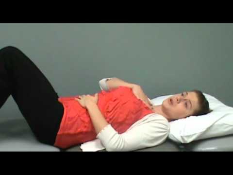
Diaphragmatic Breathing (Abdominal Breathing) Instruction and Clinical Application
Phase 2: Progressive Mobility, Motor Control & Targeted Strengthening: Phase 2 Progressive Mobilization & Motor Control: Phase 2 — Ribcage / Thoracic Compliance & Postural Expansion Phase: Because the underlying lung parenchyma is stiff, keeping the overlying thoracic cage, intercostals, and shoulder girdle as supple as possible reduces the work of breathing! Perform gentle Thoracic Expansion Exercises with arm elevation, scapular retraction, and neck/pectoral myofascial stretching.
Step 2: Restoring Flexibility & Building Supporting Muscle Strength: How to Calm the Dry, Ticklish Cough: Taking a sudden, huge deep breath stretches the stiff lung tissue and triggers a dry coughing fit! Instead, take moderate, smooth nasal breaths, sip room-temperature water, or swallow/breathe gently through pursed lips when you feel a tickle.
Clinical Assessment, Goniometry & Special Test Videos
Displayed only in Healthcare Professional mode (hidden in Patient mode)

Systematic Respiratory Physical Examination (Inspection, Palpation, Percussion, Auscultation)
Goniometric ROM & Postural / Kinematic Analysis: Pulmonary Function Testing: Restrictive pattern (FVC <80% predicted, TLC <80%, FEV1/FVC ≥0.75–0.80) and Diffusing Capacity of the Lung for Carbon Monoxide (DLCO % predicted).

Why and How Is the 6-Minute Walk Test (6MWT) Performed? Functional Capacity Assessment
Special Clinical Provocation & Neurological/Functional Tests: 6-Minute Walk Test (6MWT) with Continuous Pulse Oximetry (measuring the nadir SpO2 and exact oxygen flow rate in L/min needed to keep SpO2 ≥88–90% during ambulation!), mMRC / Borg Dyspnea Scale, and Chest Wall / Ribcage Compliance.