Exercise Intolerance and DeconditioningRapid Fatigue & Breathlessness on Exertion (Exercise Intolerance)
Exercise Intolerance and Deconditioning — Overview
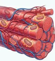
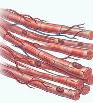
Exercise Intolerance and Cardiopulmonary Deconditioning is defined as the inability to perform physical activity at the intensity or duration expected for age and body size due to premature dyspnea (breathlessness) and/or skeletal muscle fatigue. Governed by the Fick Equation ($VO_2 = Q \times C(a-v)O_2$, where $Q = HR \times SV$), prolonged bed rest, hospitalization, post-viral syndromes, or chronic cardiopulmonary disease triggers a rapid Deconditioning Spiral: 1) Plasma volume & stroke volume drop within 48–72 hours (causing orthostatic and exertional tachycardia), 2) Skeletal muscle capillary density and mitochondrial oxidative enzymes drop by 20–30% within 2–3 weeks, and 3) Early peripheral anaerobic glycolysis produces lactic acid, which releases extra $CO_2$ through bicarbonate buffering and stimulates the brainstem respiratory center into hyperventilation and dyspnea!
Exercise intolerance and deconditioning (rapid fatigue and breathlessness on exertion) happens after an illness, hospital stay, or long period of inactivity when the heart's stroke volume and the energy factories (mitochondria) inside your leg muscles temporarily shrink. The fascinating medical truth is that half of 'shortness of breath' when walking actually comes from out-of-shape leg muscles sending 'tired' signals to your breathing center! By rebuilding your leg muscles and walking in short, comfortable intervals, both your heart rate and your breathing calm down dramatically!
Primary Causes & Biomechanics
Key Points You Should Know
- •Central limitation (reduced stroke volume / cardiac output or ventilatory restriction) combined with Peripheral Limitation (skeletal muscle atrophy, capillary rarefaction, and mitochondrial dysfunction)
- •The Lactic Acid -> Dyspnea Connection: Weak, deconditioned thigh muscles cross the Anaerobic (Ventilatory) Threshold even during slow walking—the resulting lactic acidosis drives excessive ventilation, making the patient feel 'out of breath' because their *legs* are out of shape!
- •Breaking the Spiral via FITT-VP Prescription: Reconditioning the peripheral muscles increases arteriovenous $O_2$ extraction, lowering heart rate and breathing rate at every submaximal workload!
- •Why Strengthening Your Legs Stops You From Getting Out of Breath: Weak thigh muscles produce fatigue acids after just a few steps, which instantly tells your brain to breathe faster! Strengthening your thighs and hips with chair sit-to-stands, bridges, and knee extensions makes your muscles act like fuel-efficient hybrid engines.
- •The 'Talk Test' Golden Rule for Pacing: Never push yourself until you are gasping or dizzy! Your ideal training zone is 3 to 4 out of 10 effort—where you feel warmly working, but can still speak a full sentence out loud without gasping.
- •Never Stop Walking Suddenly (The 5-Minute Cool-Down Rule): During exercise, your leg muscles pump blood back up to your heart. If you stop dead in your tracks, blood pools in your legs and causes dizziness—always finish with 3–5 minutes of slow strolling and ankle pumps!
Assessment Methods
1. Goniometric ROM & Postural / Kinematic Analysis
Cardiopulmonary Exercise Testing (CPET — VO2peak, Anaerobic Threshold, VE/VCO2 slope) or 6-Minute Walk Test (6MWT) & 1-Minute / 30-Second Sit-to-Stand Test.
2. Special Clinical Provocation & Neurological/Functional Tests
Modified Borg RPE / Dyspnea Scale (0–10), Resting -> Peak Heart Rate & Blood Pressure Response, Heart Rate Recovery at 1 Minute post-exercise (HRR-1 <12 bpm indicates autonomic deconditioning), and Pulse Oximetry (SpO2).
3. Standardized Clinical Outcome Scales & Imaging Criteria
6MWT Distance (m), 30-Second Sit-to-Stand reps, Borg RPE (target 3–4 / 10 moderate), and Heart Rate Reserve (Karvonen method).
1. Range of Motion & Posture Check
Your physiotherapist gently evaluates how comfortably and smoothly your cardiopulmonary oxygen transport cascade (Fick Equation: VO2 = Cardiac Output x Arterio-Venous O2 Difference) and peripheral skeletal muscle mitochondria moves and checks your overall posture.
2. Muscle Strength & Functional Movement Check
Your muscle balance, walking or reaching comfort, and specific gentle movement tests are evaluated without triggering sharp pain.
3. Typical Symptoms & Daily Impact Review
Disproportionate Breathlessness and Leg Heaviness during routine daily tasks (climbing one flight of stairs, carrying groceries, or walking a few blocks).
Treatment Options & Phase-Based Protocol
✨ Enriched GIF Exercise & Precaution GuidePhase 1: Acute Symptom Modulation, Tissue Protection & Early Activation
- •Phase 1 Clinical Focus & Tissue Protection: Phase 1 — Autonomic Breathing Control, Circulation & Bed/Chair Deconditioning Reversal Phase: Diaphragmatic & Pursed-Lip Breathing (slowing respiratory rate and boosting vagal tone), rhythmic ankle pumps, unloaded supine heel slides, and isometric quadriceps setting.
- •Pain, Edema & Neuromuscular Modulation: Targeted physical modalities, relative load management, and early protected mobility of the cardiopulmonary oxygen transport cascade (Fick Equation: VO2 = Cardiac Output x Arterio-Venous O2 Difference) and peripheral skeletal muscle mitochondria without provoking mechanical or inflammatory irritation.
- •Proximal & Distal Kinetic Chain Maintenance: Maintain active mobility and circulation in adjacent joints while protecting the primary healing structures in Exercise Intolerance.
Phase 2: Progressive Mobility, Motor Control & Targeted Strengthening
- •Phase 2 Progressive Mobilization & Motor Control: Phase 2 — Peripheral Large-Muscle Reconditioning Phase (Lowering Ventilatory Demand!): Train the major lower-extremity muscle groups (supine pelvic bridging, seated terminal knee extension, side-lying/standing hip abduction, and paced chair sit-to-stand—exhaling on the upward lift!) with 1–2 min recovery intervals.
- •Biomechanical Alignment & Co-Contraction: Restore balanced agonist-antagonist force couples, joint centration, and multi-planar neuromuscular control across the cardiopulmonary oxygen transport cascade (Fick Equation: VO2 = Cardiac Output x Arterio-Venous O2 Difference) and peripheral skeletal muscle mitochondria.
- •Myofascial Extensibility & Endurance: Address secondary compensatory tightness and build local muscular endurance within safe, symptom-free biomechanical parameters.
Phase 3: Functional Integration, Proprioception & Long-Term Prevention
- •Phase 3 Functional Integration & Long-Term Prevention: Phase 3 — Graded FITT-VP Aerobic Progression Phase: Frequency: 3–5 days/week; Intensity: Karvonen 40–60% Heart Rate Reserve or Borg 3–4/10 ('Talk Test' — able to speak a full sentence comfortably without gasping!); Time: start with 3 x 8–10 min bouts and build by 10%/week to 30–40 min continuous walking/cycling + 5-min cool-down (ankle pumps & calf stretches to prevent post-exercise blood pooling!).
- •Proprioceptive Balance & Dynamic Loading: Progress to task-specific closed-chain, balance, and functional movement patterns tailored to daily life, occupational, or athletic demands.
- •Self-Management & Relapse Prophylaxis: Establish lifelong ergonomic habits, home exercise maintenance, and early warning-sign awareness to prevent recurrence.
Step 1: Calming Pain, Protecting the Area & Gentle Starter Motion
- •Why Strengthening Your Legs Stops You From Getting Out of Breath: Weak thigh muscles produce fatigue acids after just a few steps, which instantly tells your brain to breathe faster! Strengthening your thighs and hips with chair sit-to-stands, bridges, and knee extensions makes your muscles act like fuel-efficient hybrid engines.
- •Calm Protection & Gentle Start: During the initial stage of Rapid Fatigue & Breathlessness on Exertion, avoid painful overloading and perform the gentle, pain-free starter movements shown below to support circulation and comfort.
- •Respecting Your Body's Signals: Every movement should stay within a comfortable, non-painful zone—never force through sharp or radiating pain.
Step 2: Restoring Flexibility & Building Supporting Muscle Strength
- •The 'Talk Test' Golden Rule for Pacing: Never push yourself until you are gasping or dizzy! Your ideal training zone is 3 to 4 out of 10 effort—where you feel warmly working, but can still speak a full sentence out loud without gasping.
- •Step-by-Step Flexibility & Support: As acute sensitivity settles, gradually build mobility and strengthen the supporting muscles around the joint at a steady, controlled pace.
- •Posture & Breathing Coordination: Keep your posture upright and breathe smoothly (never hold your breath!) during every repetition.
Step 3: Safe Daily Habits, Stamina & Preventing Recurrence
- •Never Stop Walking Suddenly (The 5-Minute Cool-Down Rule): During exercise, your leg muscles pump blood back up to your heart. If you stop dead in your tracks, blood pools in your legs and causes dizziness—always finish with 3–5 minutes of slow strolling and ankle pumps!
- •Confident Return to Daily Life: Integrate your stronger muscles and improved balance into walking, stairs, work, and recreational activities.
- •Preventing Future Flare-Ups: Continue your core protective habits and key maintenance exercises even after symptoms resolve.
Animated (GIF) Therapeutic Exercise Guide
Animated clinical exercise protocol with biomechanical angles, target muscle groups, and dosage parameters (Click image to enlarge)
Step-by-step animated exercise demonstrations you can safely perform at home (Click image to enlarge)
 🔍 Enlarge
1. Diaphragmatic & Pursed-Lip Breathing (PLB — Autonomic & Ventilatory Control)
1. Calm Belly Breathing with Pursed-Lip Exhale
Slow nasal inhalation expanding the lower ribcage and abdomen (while relaxing accessory neck/shoulder muscles), followed by prolonged expiration through pursed lips at a 1:2 ratio. Prevents dynamic airway collapse, improves basal ventilation, and reduces sympathetic tone. Specifically indicated in Exercise Intolerance rehabilitation to optimize cardiopulmonary oxygen transport cascade (Fick Equation: VO2 = Cardiac Output x Arterio-Venous O2 Difference) and peripheral skeletal muscle mitochondria biomechanics and neuromuscular control.
Sit comfortably and place one hand on your chest and the other on your belly. Breathe in slowly through your nose for 2–3 seconds so your belly gently rises, then breathe out slowly for 4–6 seconds through pursed lips as if flickering a candle. Perform smoothly within a comfortable, pain-free range to support your Rapid Fatigue & Breathlessness on Exertion recovery.
 🔍 Enlarge
2. Supine Active-Assisted Heel Slide & Synovial Diffusion Mobilization
2. Supine Heel Slide (Gentle Knee & Hip Bending)
Controlled flexion and extension of the lower extremity by sliding the heel along the support surface without axial weight-bearing. Stimulates synovial fluid diffusion across avascular cartilage and prevents capsular adhesions. Specifically indicated in Exercise Intolerance rehabilitation to optimize cardiopulmonary oxygen transport cascade (Fick Equation: VO2 = Cardiac Output x Arterio-Venous O2 Difference) and peripheral skeletal muscle mitochondria biomechanics and neuromuscular control.
While lying on your back, slowly slide your heel along the bed sheet toward your hip within a comfortable, pain-free angle, hold for 3 seconds, and gently slide it back out straight. Perform smoothly within a comfortable, pain-free range to support your Rapid Fatigue & Breathlessness on Exertion recovery.
 🔍 Enlarge
3. Seated Terminal Knee Extension (Open/Closed Chain VMO Focus)
3. Seated Knee Straightening & Hold
Concentric extension of the knee to the final 30°–0° terminal arc with isometric hold and ankle dorsiflexion. Restores terminal knee extension torque and stabilizes dynamic patellofemoral tracking. Specifically indicated in Exercise Intolerance rehabilitation to optimize cardiopulmonary oxygen transport cascade (Fick Equation: VO2 = Cardiac Output x Arterio-Venous O2 Difference) and peripheral skeletal muscle mitochondria biomechanics and neuromuscular control.
Sit upright in a chair and slowly straighten your knee completely while pulling your toes toward you. Feel the front thigh muscle tighten, hold for 5 seconds, and slowly lower your foot. Perform smoothly within a comfortable, pain-free range to support your Rapid Fatigue & Breathlessness on Exertion recovery.
 🔍 Enlarge
4. Functional Closed-Kinetic-Chain Chair Transfer (Sit-to-Stand Biomechanics)
4. Controlled Chair Sit-to-Stand Transfer
Controlled transition from sitting to standing and eccentric lowering back to the chair with symmetrical weight distribution, anterior trunk inclination ('nose over toes'), and neutral frontal knee alignment. Specifically indicated in Exercise Intolerance rehabilitation to optimize cardiopulmonary oxygen transport cascade (Fick Equation: VO2 = Cardiac Output x Arterio-Venous O2 Difference) and peripheral skeletal muscle mitochondria biomechanics and neuromuscular control.
Sit near the front of a sturdy chair with your feet hip-width apart. Lean slightly forward and push evenly through both heels to stand up tall, then slowly lower yourself back down over 3 seconds. Perform smoothly within a comfortable, pain-free range to support your Rapid Fatigue & Breathlessness on Exertion recovery.
 🔍 Enlarge
5. Rhythmic Active Ankle Dorsiflexion & Plantarflexion Pumping (Venous/Edema Pump)
5. Ankle Toe-Pulls and Pointing (Circulation & Swelling Pump)
Rhythmic alternating contraction of the tibialis anterior and gastrocnemius-soleus complex. Activates the deep venous calf pump to prevent deep vein thrombosis (DVT), clears distal edema, and maintains talocrural mobility. Specifically indicated in Exercise Intolerance rehabilitation to optimize cardiopulmonary oxygen transport cascade (Fick Equation: VO2 = Cardiac Output x Arterio-Venous O2 Difference) and peripheral skeletal muscle mitochondria biomechanics and neuromuscular control.
While lying down or sitting with your legs supported, pull your toes and feet firmly upward toward your shins, hold for 3 seconds, then point them downward like pressing a gas pedal. Perform smoothly within a comfortable, pain-free range to support your Rapid Fatigue & Breathlessness on Exertion recovery.
 🔍 Enlarge
6. Supine Lumbopelvic Bridging (Gluteus Maximus & Posterior Chain Co-Contraction)
6. Supine Hip Bridge (Controlled Pelvic Lift)
From hook-lying with transversus abdominis pre-activation, elevating the pelvis until hips, pelvis, and shoulders form a neutral diagonal line. Strengthens the gluteus maximus, hamstrings, and erector spinae synergy without lumbar hyperlordosis. Specifically indicated in Exercise Intolerance rehabilitation to optimize cardiopulmonary oxygen transport cascade (Fick Equation: VO2 = Cardiac Output x Arterio-Venous O2 Difference) and peripheral skeletal muscle mitochondria biomechanics and neuromuscular control.
Lie on your back with your knees bent and feet flat hip-width apart. Gently tighten your tummy and glutes, lift your hips until your body forms a straight line from shoulders to knees, hold for 5 seconds, and lower slowly. Perform smoothly within a comfortable, pain-free range to support your Rapid Fatigue & Breathlessness on Exertion recovery.
Movements to Avoid & Strict Precautions
1. The 'All-or-Nothing' Weekend Warrior Mistake (Going From Zero Activity to an Exhausting 1-Hour Workout!)
1. Trying to do a hard, exhausting workout on Day 1 (start with 10-minute comfortable walks and build by 10% per week!)
2. Stopping Abruptly After Brisk Walking or Cycling Without a 5-Minute Cool-Down (Causes Dizziness / Blood Pooling!)
2. Sitting or standing dead still immediately after brisk exercise without cooling down
3. Holding Your Breath During Chair Stand-Ups or Resistance Exercises
3. Holding your breath when standing up from a chair or lifting your legs
4. Exercising Immediately After a Heavy Meal or in Extreme Heat/Humidity
4. Exercising right after a large meal (wait 1.5–2 hours, as digestion diverts blood flow away from muscles!)
Exercise Intolerance and Deconditioning — Video Guide
Click directly on any video to open and watch in a window, or review details via 📋 Summary & Options.
Condition Overview Video (1 Video)
Summary of pathophysiology, biomechanics, and clinical presentation
What the condition is, why it develops, and its basic mechanism

What Are Exercise Intolerance and Deconditioning? Causes and Physiology
Exercise Intolerance and Cardiopulmonary Deconditioning is defined as the inability to perform physical activity at the intensity or duration expected for age and body size due to premature dyspnea (breathlessness) and/or skeletal muscle fatigue. Governed by the Fick Equation ($VO_2 = Q \times C(a-v)O_2$, where $Q = HR \times SV$), prolonged bed rest, hospitalization, post-viral syndromes, or chronic cardiopulmonary disease triggers a rapid Deconditioning Spiral: 1) Plasma volume & stroke volume drop within 48–72 hours (causing orthostatic and exertional tachycardia), 2) Skeletal muscle capillary density and mitochondrial oxidative enzymes drop by 20–30% within 2–3 weeks, and 3) Early peripheral anaerobic glycolysis produces lactic acid, which releases extra $CO_2$ through bicarbonate buffering and stimulates the brainstem respiratory center into hyperventilation and dyspnea!
Exercise intolerance and deconditioning (rapid fatigue and breathlessness on exertion) happens after an illness, hospital stay, or long period of inactivity when the heart's stroke volume and the energy factories (mitochondria) inside your leg muscles temporarily shrink. The fascinating medical truth is that half of 'shortness of breath' when walking actually comes from out-of-shape leg muscles sending 'tired' signals to your breathing center! By rebuilding your leg muscles and walking in short, comfortable intervals, both your heart rate and your breathing calm down dramatically!
Treatment and Exercise Videos
Phase-based physiotherapy protocols, mobilization, and therapeutic exercises
Step-by-step recovery and safe home exercise applications

Safe Aerobic Exercise Guide for Heart Health and Functional Capacity
Phase 1: Acute Symptom Modulation, Tissue Protection & Early Activation: Phase 1 Clinical Focus & Tissue Protection: Phase 1 — Autonomic Breathing Control, Circulation & Bed/Chair Deconditioning Reversal Phase: Diaphragmatic & Pursed-Lip Breathing (slowing respiratory rate and boosting vagal tone), rhythmic ankle pumps, unloaded supine heel slides, and isometric quadriceps setting.
Step 1: Calming Pain, Protecting the Area & Gentle Starter Motion: Why Strengthening Your Legs Stops You From Getting Out of Breath: Weak thigh muscles produce fatigue acids after just a few steps, which instantly tells your brain to breathe faster! Strengthening your thighs and hips with chair sit-to-stands, bridges, and knee extensions makes your muscles act like fuel-efficient hybrid engines.

Graded Exercise Training in Cardiac Rehabilitation and Exercise Intolerance
Phase 2: Progressive Mobility, Motor Control & Targeted Strengthening: Phase 2 Progressive Mobilization & Motor Control: Phase 2 — Peripheral Large-Muscle Reconditioning Phase (Lowering Ventilatory Demand!): Train the major lower-extremity muscle groups (supine pelvic bridging, seated terminal knee extension, side-lying/standing hip abduction, and paced chair sit-to-stand—exhaling on the upward lift!) with 1–2 min recovery intervals.
Step 2: Restoring Flexibility & Building Supporting Muscle Strength: The 'Talk Test' Golden Rule for Pacing: Never push yourself until you are gasping or dizzy! Your ideal training zone is 3 to 4 out of 10 effort—where you feel warmly working, but can still speak a full sentence out loud without gasping.
Clinical Assessment, Goniometry & Special Test Videos
Displayed only in Healthcare Professional mode (hidden in Patient mode)

Why and How Is the 6-Minute Walk Test (6MWT) Performed? Functional Capacity Assessment
Goniometric ROM & Postural / Kinematic Analysis: Cardiopulmonary Exercise Testing (CPET — VO2peak, Anaerobic Threshold, VE/VCO2 slope) or 6-Minute Walk Test (6MWT) & 1-Minute / 30-Second Sit-to-Stand Test.

Cardiopulmonary Exercise Testing (CPET) and VO2max Capacity Evaluation
Special Clinical Provocation & Neurological/Functional Tests: Modified Borg RPE / Dyspnea Scale (0–10), Resting -> Peak Heart Rate & Blood Pressure Response, Heart Rate Recovery at 1 Minute post-exercise (HRR-1 <12 bpm indicates autonomic deconditioning), and Pulse Oximetry (SpO2).