Coronary Artery Disease (CAD)Heart Artery Blockage / Narrowing (Coronary Artery Disease)
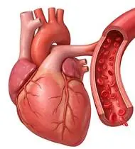
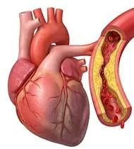
Coronary Artery Disease (CAD) — Overview
Coronary Artery Disease (CAD / Ischemic Heart Disease) is caused by atherosclerotic plaque stenosis or thrombosis in the epicardial coronary arteries, creating an imbalance between myocardial oxygen supply and myocardial oxygen demand (quantified clinically by the Rate-Pressure Product [RPP = Heart Rate x Systolic Blood Pressure]). Following acute Myocardial Infarction (MI), Percutaneous Coronary Intervention (PCI / Stent), or Coronary Artery Bypass Grafting (CABG), structured Phase I to Phase IV Cardiac Rehabilitation is a Class IA life-saving medical intervention proven to reduce cardiovascular mortality by 26% and hospital readmissions by 31%! Cardiac rehab works by lowering the Rate-Pressure Product (HR x SBP) at any given submaximal workload, raising the anginal threshold, improving coronary endothelial nitric oxide function, and stabilizing autonomic heart rate variability!
Coronary Artery Disease (CAD / heart artery narrowing, stent, or bypass recovery) occurs when the arteries feeding oxygen to the heart muscle become narrowed or treated with a stent or bypass surgery (CABG). Cardiac Rehabilitation is one of the most powerful medicines in cardiology: regular, properly paced walking and rhythmic leg exercises train your body's muscles to use oxygen so efficiently that your heart beats slower and at lower blood pressure during everyday life—cutting future heart risks by over 25%!
Primary Causes & Biomechanics
Key Points You Should Know
- •Coronary endothelial dysfunction and atherosclerotic stenosis limiting coronary blood flow reserve during exertion
- •Myocardial Oxygen Demand Law ($RPP = HR \times SBP$): Upper-extremity overhead exercise and isometric breath-holding (Valsalva) disproportionately spike Systolic Blood Pressure and Rate-Pressure Product compared to rhythmic lower-extremity walking/cycling!
- •Post-CABG Sternotomy Protection Rule: After median sternotomy bypass surgery, strict Sternal Precautions ('Keep Your Move in the Tube' — no unilateral pulling, no pushing >2–4 kg, no bilateral arms behind back) apply for 6–8 weeks while the sternum unites!
- •Why a 10-Minute Warm-Up and 10-Minute Cool-Down Are Non-Negotiable for the Heart: Starting exercise abruptly before the heart's own coronary arteries have dilated can starve the heart muscle of oxygen, while stopping abruptly lets blood pool in the legs and triggers rhythm changes. Always spend 5–10 minutes warming up slowly and 5–10 minutes strolling to cool down!
- •If You Take Beta-Blocker Medication, Use the 'Talk Test' (Borg 11–13): Heart medications (like metoprolol, bisoprolol, or carvedilol) intentionally keep your heart rate low and steady. Instead of chasing a high heart-rate number, exercise at a comfortable, rhythmic pace where you can speak a full sentence easily without gasping!
- •After Bypass Surgery (CABG) — 'Keep Your Arms in the Tube' for 6–8 Weeks: While your breastbone (sternum) heals for the first 6–8 weeks, keep your upper arms tucked close to your ribs (as if inside a narrow tube)—never push yourself up from a chair with one arm, reach behind your back, or lift more than a 2-liter bottle!
Assessment Methods
1. Goniometric ROM & Postural / Kinematic Analysis
Symptom-Limited Exercise Tolerance Test (ETT / ECG Stress Test), 6-Minute Walk Test (6MWT), Karvonen Heart Rate Reserve ($HR_{target} = HR_{rest} + 40 ext{--}70\% \times [HR_{max} - HR_{rest}]$, or $HR_{rest} + 20 ext{--}30 ext{ bpm}$ if on Beta-Blockers!), and Borg RPE Scale (6–20: target 11–13 'Fairly Light to Somewhat Hard' / 0–10: target 3–4).
2. Special Clinical Provocation & Neurological/Functional Tests
Resting and exercise Heart Rate, Blood Pressure, SpO2, Angina Scale (0–4), and Sternal Stability Check (in post-CABG patients).
3. Standardized Clinical Outcome Scales & Imaging Criteria
MET Capacity (1 MET = 3.5 mL O2/kg/min; target ≥5–8 METs for full daily independence), 6MWT, Borg RPE (11–13), and LVEF (%).
1. Range of Motion & Posture Check
Your physiotherapist gently evaluates how comfortably and smoothly your coronary arteries (LAD, LCx, RCA atherosclerotic plaques), myocardium, and autonomic cardiovascular regulation moves and checks your overall posture.
2. Muscle Strength & Functional Movement Check
Your muscle balance, walking or reaching comfort, and specific gentle movement tests are evaluated without triggering sharp pain.
3. Typical Symptoms & Daily Impact Review
Stable Exertional Angina (In Untreated/Residual Stenosis): A feeling of pressure, tightness, squeezing, or heaviness in the center of the chest (sometimes radiating to the left shoulder/arm, neck, jaw, or back) triggered by physical exertion, cold air, or heavy meals, and relieved within 3–5 minutes by rest or sublingual nitroglycerin.
Treatment Options & Phase-Based Protocol
✨ Enriched GIF Exercise & Precaution GuidePhase 1: Acute Symptom Modulation, Tissue Protection & Early Activation
- •Phase 1 Clinical Focus & Tissue Protection: Phase I — Inpatient / Early Mobilization Phase (1–3 METs): Diaphragmatic breathing (splinting the chest with a pillow if post-CABG!), hourly ankle pumps (especially in the saphenous vein graft leg!), unloaded supine heel slides, and seated-to-hallway walking keeping HR within resting HR + 20 bpm.
- •Pain, Edema & Neuromuscular Modulation: Targeted physical modalities, relative load management, and early protected mobility of the coronary arteries (LAD, LCx, RCA atherosclerotic plaques), myocardium, and autonomic cardiovascular regulation without provoking mechanical or inflammatory irritation.
- •Proximal & Distal Kinetic Chain Maintenance: Maintain active mobility and circulation in adjacent joints while protecting the primary healing structures in Coronary Artery Disease.
Phase 2: Progressive Mobility, Motor Control & Targeted Strengthening
- •Phase 2 Progressive Mobilization & Motor Control: Phase II–III — Monitored Outpatient Aerobic & Rhythmic Lower-Limb Phase (Weeks 2–12): Mandatory 5–10 min Warm-Up (allows coronary vasodilation and prevents ischemic ST-depression!) -> 20–40 min rhythmic aerobic walking or stationary cycling at Borg RPE 11–13 (Talk Test comfortable) -> Rhythmic non-Valsalva lower-limb exercises (chair sit-to-stand, wall mini-squats, heel raises, pelvic bridges — counting out loud on every rep!) -> Mandatory 5–10 min Cool-Down (prevents post-exercise hypotension and catecholamine-induced arrhythmias!).
- •Biomechanical Alignment & Co-Contraction: Restore balanced agonist-antagonist force couples, joint centration, and multi-planar neuromuscular control across the coronary arteries (LAD, LCx, RCA atherosclerotic plaques), myocardium, and autonomic cardiovascular regulation.
- •Myofascial Extensibility & Endurance: Address secondary compensatory tightness and build local muscular endurance within safe, symptom-free biomechanical parameters.
Phase 3: Functional Integration, Proprioception & Long-Term Prevention
- •Phase 3 Functional Integration & Long-Term Prevention: Phase IV — Lifelong Secondary Prevention & Scapular/Thoracic Phase: Scapular retraction & thoracic expansion (restoring chest wall mobility once the sternum is healed at 6–8 weeks post-CABG), lifelong 150+ min/week aerobic activity, and risk-factor control.
- •Proprioceptive Balance & Dynamic Loading: Progress to task-specific closed-chain, balance, and functional movement patterns tailored to daily life, occupational, or athletic demands.
- •Self-Management & Relapse Prophylaxis: Establish lifelong ergonomic habits, home exercise maintenance, and early warning-sign awareness to prevent recurrence.
Step 1: Calming Pain, Protecting the Area & Gentle Starter Motion
- •Why a 10-Minute Warm-Up and 10-Minute Cool-Down Are Non-Negotiable for the Heart: Starting exercise abruptly before the heart's own coronary arteries have dilated can starve the heart muscle of oxygen, while stopping abruptly lets blood pool in the legs and triggers rhythm changes. Always spend 5–10 minutes warming up slowly and 5–10 minutes strolling to cool down!
- •Calm Protection & Gentle Start: During the initial stage of Heart Artery Blockage / Narrowing, avoid painful overloading and perform the gentle, pain-free starter movements shown below to support circulation and comfort.
- •Respecting Your Body's Signals: Every movement should stay within a comfortable, non-painful zone—never force through sharp or radiating pain.
Step 2: Restoring Flexibility & Building Supporting Muscle Strength
- •If You Take Beta-Blocker Medication, Use the 'Talk Test' (Borg 11–13): Heart medications (like metoprolol, bisoprolol, or carvedilol) intentionally keep your heart rate low and steady. Instead of chasing a high heart-rate number, exercise at a comfortable, rhythmic pace where you can speak a full sentence easily without gasping!
- •Step-by-Step Flexibility & Support: As acute sensitivity settles, gradually build mobility and strengthen the supporting muscles around the joint at a steady, controlled pace.
- •Posture & Breathing Coordination: Keep your posture upright and breathe smoothly (never hold your breath!) during every repetition.
Step 3: Safe Daily Habits, Stamina & Preventing Recurrence
- •After Bypass Surgery (CABG) — 'Keep Your Arms in the Tube' for 6–8 Weeks: While your breastbone (sternum) heals for the first 6–8 weeks, keep your upper arms tucked close to your ribs (as if inside a narrow tube)—never push yourself up from a chair with one arm, reach behind your back, or lift more than a 2-liter bottle!
- •Confident Return to Daily Life: Integrate your stronger muscles and improved balance into walking, stairs, work, and recreational activities.
- •Preventing Future Flare-Ups: Continue your core protective habits and key maintenance exercises even after symptoms resolve.
Animated (GIF) Therapeutic Exercise Guide
Animated clinical exercise protocol with biomechanical angles, target muscle groups, and dosage parameters (Click image to enlarge)
Step-by-step animated exercise demonstrations you can safely perform at home (Click image to enlarge)
 🔍 Enlarge
1. Rhythmic Active Ankle Dorsiflexion & Plantarflexion Pumping (Venous/Edema Pump)
1. Ankle Toe-Pulls and Pointing (Circulation & Swelling Pump)
Rhythmic alternating contraction of the tibialis anterior and gastrocnemius-soleus complex. Activates the deep venous calf pump to prevent deep vein thrombosis (DVT), clears distal edema, and maintains talocrural mobility. Specifically indicated in Coronary Artery Disease rehabilitation to optimize coronary arteries (LAD, LCx, RCA atherosclerotic plaques), myocardium, and autonomic cardiovascular regulation biomechanics and neuromuscular control.
While lying down or sitting with your legs supported, pull your toes and feet firmly upward toward your shins, hold for 3 seconds, then point them downward like pressing a gas pedal. Perform smoothly within a comfortable, pain-free range to support your Heart Artery Blockage / Narrowing recovery.
 🔍 Enlarge
2. Thoracic Expansion Exercise (TEE) with Bilateral Arm Elevation & 3s Inspiratory Hold
2. Chest-Opening Deep Breath with Arm Raise
Synchronizing deep nasal inspiration with bilateral shoulder flexion/abduction and a 3-second end-inspiratory hold before passive/pursed-lip expiration. Opens collateral ventilation channels to prevent basal atelectasis and maintains costovertebral compliance. Specifically indicated in Coronary Artery Disease rehabilitation to optimize coronary arteries (LAD, LCx, RCA atherosclerotic plaques), myocardium, and autonomic cardiovascular regulation biomechanics and neuromuscular control.
While sitting upright, breathe in deeply through your nose while opening your arms upward and outward to expand your ribcage. Hold the breath inside for 3 seconds, then exhale slowly while lowering your arms. Perform smoothly within a comfortable, pain-free range to support your Heart Artery Blockage / Narrowing recovery.
 🔍 Enlarge
3. Seated Terminal Knee Extension (Open/Closed Chain VMO Focus)
3. Seated Knee Straightening & Hold
Concentric extension of the knee to the final 30°–0° terminal arc with isometric hold and ankle dorsiflexion. Restores terminal knee extension torque and stabilizes dynamic patellofemoral tracking. Specifically indicated in Coronary Artery Disease rehabilitation to optimize coronary arteries (LAD, LCx, RCA atherosclerotic plaques), myocardium, and autonomic cardiovascular regulation biomechanics and neuromuscular control.
Sit upright in a chair and slowly straighten your knee completely while pulling your toes toward you. Feel the front thigh muscle tighten, hold for 5 seconds, and slowly lower your foot. Perform smoothly within a comfortable, pain-free range to support your Heart Artery Blockage / Narrowing recovery.
 🔍 Enlarge
4. Functional Closed-Kinetic-Chain Chair Transfer (Sit-to-Stand Biomechanics)
4. Controlled Chair Sit-to-Stand Transfer
Controlled transition from sitting to standing and eccentric lowering back to the chair with symmetrical weight distribution, anterior trunk inclination ('nose over toes'), and neutral frontal knee alignment. Specifically indicated in Coronary Artery Disease rehabilitation to optimize coronary arteries (LAD, LCx, RCA atherosclerotic plaques), myocardium, and autonomic cardiovascular regulation biomechanics and neuromuscular control.
Sit near the front of a sturdy chair with your feet hip-width apart. Lean slightly forward and push evenly through both heels to stand up tall, then slowly lower yourself back down over 3 seconds. Perform smoothly within a comfortable, pain-free range to support your Heart Artery Blockage / Narrowing recovery.
 🔍 Enlarge
5. Diaphragmatic & Pursed-Lip Breathing (PLB — Autonomic & Ventilatory Control)
5. Calm Belly Breathing with Pursed-Lip Exhale
Slow nasal inhalation expanding the lower ribcage and abdomen (while relaxing accessory neck/shoulder muscles), followed by prolonged expiration through pursed lips at a 1:2 ratio. Prevents dynamic airway collapse, improves basal ventilation, and reduces sympathetic tone. Specifically indicated in Coronary Artery Disease rehabilitation to optimize coronary arteries (LAD, LCx, RCA atherosclerotic plaques), myocardium, and autonomic cardiovascular regulation biomechanics and neuromuscular control.
Sit comfortably and place one hand on your chest and the other on your belly. Breathe in slowly through your nose for 2–3 seconds so your belly gently rises, then breathe out slowly for 4–6 seconds through pursed lips as if flickering a candle. Perform smoothly within a comfortable, pain-free range to support your Heart Artery Blockage / Narrowing recovery.
 🔍 Enlarge
6. Supine Active-Assisted Heel Slide & Synovial Diffusion Mobilization
6. Supine Heel Slide (Gentle Knee & Hip Bending)
Controlled flexion and extension of the lower extremity by sliding the heel along the support surface without axial weight-bearing. Stimulates synovial fluid diffusion across avascular cartilage and prevents capsular adhesions. Specifically indicated in Coronary Artery Disease rehabilitation to optimize coronary arteries (LAD, LCx, RCA atherosclerotic plaques), myocardium, and autonomic cardiovascular regulation biomechanics and neuromuscular control.
While lying on your back, slowly slide your heel along the bed sheet toward your hip within a comfortable, pain-free angle, hold for 3 seconds, and gently slide it back out straight. Perform smoothly within a comfortable, pain-free range to support your Heart Artery Blockage / Narrowing recovery.
Movements to Avoid & Strict Precautions
1. Isometric Straining, Planks, Heavy Lifting, or Breath-Holding (Valsalva Spikes Blood Pressure & Heart Wall Stress!)
1. NEVER hold your breath while exercising or lifting, and avoid planks, push-ups, or heavy weights (always count out loud!)
2. Shoveling Snow or Exercising in Freezing Cold Wind (Cold Air + Overhead Shoveling Causes Coronary Artery Spasm!)
2. Shoveling snow or exercising outdoors in freezing wind (cold constricts coronary arteries while arm lifting spikes blood pressure!)
3. Skipping the 5–10 Minute Warm-Up or Stopping Abruptly Without a Cool-Down
3. Starting fast exercise without a gradual warm-up or sitting down immediately after fast walking
4. Pushing Up With One Arm or Pulling Heavy Doors in the First 6–8 Weeks After Bypass Surgery (Sternotomy)
4. Using one arm to push out of bed/chairs or lifting >2–3 kg during the first 6–8 weeks after open-heart bypass surgery
Coronary Artery Disease (CAD) — Video Guide
Click directly on any video to open and watch in a window, or review details via 📋 Summary & Options.
Condition Overview Video (1 Video)
Summary of pathophysiology, biomechanics, and clinical presentation
What the condition is, why it develops, and its basic mechanism

What Are Coronary Artery Disease (CAD) and Myocardial Infarction? Symptoms & Risks
Coronary Artery Disease (CAD / Ischemic Heart Disease) is caused by atherosclerotic plaque stenosis or thrombosis in the epicardial coronary arteries, creating an imbalance between myocardial oxygen supply and myocardial oxygen demand (quantified clinically by the Rate-Pressure Product [RPP = Heart Rate x Systolic Blood Pressure]). Following acute Myocardial Infarction (MI), Percutaneous Coronary Intervention (PCI / Stent), or Coronary Artery Bypass Grafting (CABG), structured Phase I to Phase IV Cardiac Rehabilitation is a Class IA life-saving medical intervention proven to reduce cardiovascular mortality by 26% and hospital readmissions by 31%! Cardiac rehab works by lowering the Rate-Pressure Product (HR x SBP) at any given submaximal workload, raising the anginal threshold, improving coronary endothelial nitric oxide function, and stabilizing autonomic heart rate variability!
Coronary Artery Disease (CAD / heart artery narrowing, stent, or bypass recovery) occurs when the arteries feeding oxygen to the heart muscle become narrowed or treated with a stent or bypass surgery (CABG). Cardiac Rehabilitation is one of the most powerful medicines in cardiology: regular, properly paced walking and rhythmic leg exercises train your body's muscles to use oxygen so efficiently that your heart beats slower and at lower blood pressure during everyday life—cutting future heart risks by over 25%!
Treatment and Exercise Videos
Phase-based physiotherapy protocols, mobilization, and therapeutic exercises
Step-by-step recovery and safe home exercise applications

Safe Aerobic Exercise Guide for Heart Health and Functional Capacity
Phase 1: Acute Symptom Modulation, Tissue Protection & Early Activation: Phase 1 Clinical Focus & Tissue Protection: Phase I — Inpatient / Early Mobilization Phase (1–3 METs): Diaphragmatic breathing (splinting the chest with a pillow if post-CABG!), hourly ankle pumps (especially in the saphenous vein graft leg!), unloaded supine heel slides, and seated-to-hallway walking keeping HR within resting HR + 20 bpm.
Step 1: Calming Pain, Protecting the Area & Gentle Starter Motion: Why a 10-Minute Warm-Up and 10-Minute Cool-Down Are Non-Negotiable for the Heart: Starting exercise abruptly before the heart's own coronary arteries have dilated can starve the heart muscle of oxygen, while stopping abruptly lets blood pool in the legs and triggers rhythm changes. Always spend 5–10 minutes warming up slowly and 5–10 minutes strolling to cool down!

Graded Exercise Training in Cardiac Rehabilitation and Exercise Intolerance
Phase 2: Progressive Mobility, Motor Control & Targeted Strengthening: Phase 2 Progressive Mobilization & Motor Control: Phase II–III — Monitored Outpatient Aerobic & Rhythmic Lower-Limb Phase (Weeks 2–12): Mandatory 5–10 min Warm-Up (allows coronary vasodilation and prevents ischemic ST-depression!) -> 20–40 min rhythmic aerobic walking or stationary cycling at Borg RPE 11–13 (Talk Test comfortable) -> Rhythmic non-Valsalva lower-limb exercises (chair sit-to-stand, wall mini-squats, heel raises, pelvic bridges — counting out loud on every rep!) -> Mandatory 5–10 min Cool-Down (prevents post-exercise hypotension and catecholamine-induced arrhythmias!).
Step 2: Restoring Flexibility & Building Supporting Muscle Strength: If You Take Beta-Blocker Medication, Use the 'Talk Test' (Borg 11–13): Heart medications (like metoprolol, bisoprolol, or carvedilol) intentionally keep your heart rate low and steady. Instead of chasing a high heart-rate number, exercise at a comfortable, rhythmic pace where you can speak a full sentence easily without gasping!
Clinical Assessment, Goniometry & Special Test Videos
Displayed only in Healthcare Professional mode (hidden in Patient mode)

Cardiopulmonary Exercise Testing (CPET) and VO2max Capacity Evaluation
Goniometric ROM & Postural / Kinematic Analysis: Symptom-Limited Exercise Tolerance Test (ETT / ECG Stress Test), 6-Minute Walk Test (6MWT), Karvonen Heart Rate Reserve ($HR_{target} = HR_{rest} + 40 ext{--}70\% \times [HR_{max} - HR_{rest}]$, or $HR_{rest} + 20 ext{--}30 ext{ bpm}$ if on Beta-Blockers!), and Borg RPE Scale (6–20: target 11–13 'Fairly Light to Somewhat Hard' / 0–10: target 3–4).

Why and How Is the 6-Minute Walk Test (6MWT) Performed? Functional Capacity Assessment
Special Clinical Provocation & Neurological/Functional Tests: Resting and exercise Heart Rate, Blood Pressure, SpO2, Angina Scale (0–4), and Sternal Stability Check (in post-CABG patients).