BronchiectasisPermanent Widening of Lung Airways (Bronchiectasis)
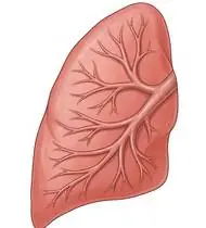
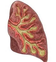
Bronchiectasis — Overview
Bronchiectasis is a chronic suppurative airway disease characterized by irreversible, abnormal dilatation and distortion of medium-sized bronchi (diagnosed via High-Resolution CT [HRCT]: broncho-arterial ratio >1.0 ['signet-ring sign'], lack of bronchial tapering, and visible bronchi within 1 cm of the pleura). It is driven by Cole's Vicious Vortex: 1) Impaired Mucociliary Clearance -> 2) Mucus Pooling & Bacterial Colonization (*H. influenzae, P. aeruginosa*) -> 3) Neutrophilic Inflammation (neutrophil elastase) -> 4) Progressive Structural Bronchial Wall Destruction. Therefore, twice-daily Airway Clearance Physiotherapy (Bronchial Hygiene) is a mandatory, lifelong cornerstone of treatment (on par with antibiotics!) to break Cole's Vicious Vortex!
Bronchiectasis (permanent widening of the lung airways) is a condition in which parts of the air tubes (bronchi) have become stretched out and widened, forming small pockets where mucus naturally collects instead of sweeping upward on its own. Because pooled mucus invites infection, your 15–20 minute morning and evening Airway Clearance Physiotherapy routine acts as your lungs' daily 'toothbrushing'—sweeping those pockets clean, preventing flare-ups, and stopping daytime coughing fits!
Primary Causes & Biomechanics
Key Points You Should Know
- •Post-infectious damage (severe childhood pneumonia, tuberculosis, pertussis), immunodeficiency, primary ciliary dyskinesia (Kartagener's), allergic bronchopulmonary aspergillosis (ABPA), or non-CF/CF genetic causes
- •Loss of Ciliary Escalator & Floppy Bronchial Walls: Destroyed elastic/cartilage walls allow mucus to pool in dependent saccular/cylindrical pockets while the bronchial walls collapse easily during forceful coughing
- •Why Huffing (Forced Expiration Technique — FET) and Oscillatory PEP are superior to violent coughing: Huffing shifts the Equal Pressure Point (EPP) peripherally without collapsing the weakened bronchiectatic walls!
- •Why 'Huffing' Works Better Than Hard Coughing: In bronchiectasis, the widened airway walls are soft; if you cough violently, the walls slam shut and trap the phlegm inside! Instead, take 3 slow, deep breaths (holding each for 3 seconds so air slips *behind* the mucus), then open your mouth in an 'O' shape and breathe out with a firm 'HAAAAF' as if fogging up a bathroom mirror. This slides the phlegm effortlessly to the top so one small cough clears it!
- •Clearing Your Lungs Morning and Evening Stops Daytime Social Coughing: Spending 15 minutes clearing your airways first thing in the morning and 2 hours before bed empties the mucus pockets so you can go through your day without embarrassing coughing spells.
- •Hydration + Brisk Walking Thins the Mucus: Drinking plenty of water and taking a daily brisk walk makes mucus watery and easy to clear.
Assessment Methods
1. Goniometric ROM & Postural / Kinematic Analysis
HRCT Lobar Localization (determining which exact lung lobes/segments contain pooled bronchiectatic secretions to guide targeted positioning!), 24-hour sputum volume & purulence (Murray-Washington color chart: mucoid white -> mucopurulent yellow -> purulent dark green), and spirometry.
2. Special Clinical Provocation & Neurological/Functional Tests
6-Minute Walk Test (6MWT), Leicester Cough Questionnaire (LCQ), mMRC dyspnea scale, and thoracic expansion symmetry.
3. Standardized Clinical Outcome Scales & Imaging Criteria
24-Hour Sputum Volume/Color, Leicester Cough Questionnaire (LCQ), 6MWT, and FEV1/FVC.
1. Range of Motion & Posture Check
Your physiotherapist gently evaluates how comfortably and smoothly your medium-sized bronchi (permanent abnormal dilatation and destruction of bronchial walls) and mucociliary clearance escalator moves and checks your overall posture.
2. Muscle Strength & Functional Movement Check
Your muscle balance, walking or reaching comfort, and specific gentle movement tests are evaluated without triggering sharp pain.
3. Typical Symptoms & Daily Impact Review
Chronic Daily Productive Cough bringing up mucoid or yellow-green sputum (especially in the morning or when changing posture).
Treatment Options & Phase-Based Protocol
✨ Enriched GIF Exercise & Precaution GuidePhase 1: Acute Symptom Modulation, Tissue Protection & Early Activation
- •Phase 1 Clinical Focus & Tissue Protection: Phase 1 — Targeted Airway Clearance (ACBT, Autogenic Drainage & OPEP) Phase (Twice Daily — Morning & Evening!): Perform nebulized hypertonic saline (3–7%)/bronchodilator BEFORE physiotherapy, followed by Active Cycle of Breathing Techniques (ACBT: 1. Breathing Control -> 2. 3–4 Thoracic Expansion Exercises with 3s inspiratory hold [filling collateral Pores of Kohn behind the mucus plug!] -> 3. Open-Glottis 'Huffing' [low-volume huff to mobilize peripheral mucus, high-volume huff to clear central mucus!]) + Oscillatory PEP devices (Flutter, Acapella, Aerobika) and Modified Postural Drainage (NEVER head-down if GERD is present!).
- •Pain, Edema & Neuromuscular Modulation: Targeted physical modalities, relative load management, and early protected mobility of the medium-sized bronchi (permanent abnormal dilatation and destruction of bronchial walls) and mucociliary clearance escalator without provoking mechanical or inflammatory irritation.
- •Proximal & Distal Kinetic Chain Maintenance: Maintain active mobility and circulation in adjacent joints while protecting the primary healing structures in Bronchiectasis.
Phase 2: Progressive Mobility, Motor Control & Targeted Strengthening
- •Phase 2 Progressive Mobilization & Motor Control: Phase 2 — Thoracic Cage Mobility & Inspiratory Muscle Phase: Bilateral arm elevation with deep costal inspiration, scapular retraction, and diaphragmatic/pursed-lip breathing.
- •Biomechanical Alignment & Co-Contraction: Restore balanced agonist-antagonist force couples, joint centration, and multi-planar neuromuscular control across the medium-sized bronchi (permanent abnormal dilatation and destruction of bronchial walls) and mucociliary clearance escalator.
- •Myofascial Extensibility & Endurance: Address secondary compensatory tightness and build local muscular endurance within safe, symptom-free biomechanical parameters.
Phase 3: Functional Integration, Proprioception & Long-Term Prevention
- •Phase 3 Functional Integration & Long-Term Prevention: Phase 3 — Aerobic Exercise (Natural Mucus Mobilizer!) & Lower-Limb Conditioning Phase: 30–40 min of brisk walking, stationary cycling, or interval exercise 4–5 days/week (increased ventilatory airflow during exercise shears mucus off bronchial walls!), plus chair sit-to-stand, squats, and heel raises.
- •Proprioceptive Balance & Dynamic Loading: Progress to task-specific closed-chain, balance, and functional movement patterns tailored to daily life, occupational, or athletic demands.
- •Self-Management & Relapse Prophylaxis: Establish lifelong ergonomic habits, home exercise maintenance, and early warning-sign awareness to prevent recurrence.
Step 1: Calming Pain, Protecting the Area & Gentle Starter Motion
- •Why 'Huffing' Works Better Than Hard Coughing: In bronchiectasis, the widened airway walls are soft; if you cough violently, the walls slam shut and trap the phlegm inside! Instead, take 3 slow, deep breaths (holding each for 3 seconds so air slips *behind* the mucus), then open your mouth in an 'O' shape and breathe out with a firm 'HAAAAF' as if fogging up a bathroom mirror. This slides the phlegm effortlessly to the top so one small cough clears it!
- •Calm Protection & Gentle Start: During the initial stage of Permanent Widening of Lung Airways, avoid painful overloading and perform the gentle, pain-free starter movements shown below to support circulation and comfort.
- •Respecting Your Body's Signals: Every movement should stay within a comfortable, non-painful zone—never force through sharp or radiating pain.
Step 2: Restoring Flexibility & Building Supporting Muscle Strength
- •Clearing Your Lungs Morning and Evening Stops Daytime Social Coughing: Spending 15 minutes clearing your airways first thing in the morning and 2 hours before bed empties the mucus pockets so you can go through your day without embarrassing coughing spells.
- •Step-by-Step Flexibility & Support: As acute sensitivity settles, gradually build mobility and strengthen the supporting muscles around the joint at a steady, controlled pace.
- •Posture & Breathing Coordination: Keep your posture upright and breathe smoothly (never hold your breath!) during every repetition.
Step 3: Safe Daily Habits, Stamina & Preventing Recurrence
- •Hydration + Brisk Walking Thins the Mucus: Drinking plenty of water and taking a daily brisk walk makes mucus watery and easy to clear.
- •Confident Return to Daily Life: Integrate your stronger muscles and improved balance into walking, stairs, work, and recreational activities.
- •Preventing Future Flare-Ups: Continue your core protective habits and key maintenance exercises even after symptoms resolve.
Animated (GIF) Therapeutic Exercise Guide
Animated clinical exercise protocol with biomechanical angles, target muscle groups, and dosage parameters (Click image to enlarge)
Step-by-step animated exercise demonstrations you can safely perform at home (Click image to enlarge)
 🔍 Enlarge
1. Diaphragmatic & Pursed-Lip Breathing (PLB — Autonomic & Ventilatory Control)
1. Calm Belly Breathing with Pursed-Lip Exhale
Slow nasal inhalation expanding the lower ribcage and abdomen (while relaxing accessory neck/shoulder muscles), followed by prolonged expiration through pursed lips at a 1:2 ratio. Prevents dynamic airway collapse, improves basal ventilation, and reduces sympathetic tone. Specifically indicated in Bronchiectasis rehabilitation to optimize medium-sized bronchi (permanent abnormal dilatation and destruction of bronchial walls) and mucociliary clearance escalator biomechanics and neuromuscular control.
Sit comfortably and place one hand on your chest and the other on your belly. Breathe in slowly through your nose for 2–3 seconds so your belly gently rises, then breathe out slowly for 4–6 seconds through pursed lips as if flickering a candle. Perform smoothly within a comfortable, pain-free range to support your Permanent Widening of Lung Airways recovery.
 🔍 Enlarge
2. Thoracic Expansion Exercise (TEE) with Bilateral Arm Elevation & 3s Inspiratory Hold
2. Chest-Opening Deep Breath with Arm Raise
Synchronizing deep nasal inspiration with bilateral shoulder flexion/abduction and a 3-second end-inspiratory hold before passive/pursed-lip expiration. Opens collateral ventilation channels to prevent basal atelectasis and maintains costovertebral compliance. Specifically indicated in Bronchiectasis rehabilitation to optimize medium-sized bronchi (permanent abnormal dilatation and destruction of bronchial walls) and mucociliary clearance escalator biomechanics and neuromuscular control.
While sitting upright, breathe in deeply through your nose while opening your arms upward and outward to expand your ribcage. Hold the breath inside for 3 seconds, then exhale slowly while lowering your arms. Perform smoothly within a comfortable, pain-free range to support your Permanent Widening of Lung Airways recovery.
 🔍 Enlarge
3. Wall-Referenced Axial Spinal Extension & Postural Decompression
3. Wall-Supported Tall Standing & Back Alignment
Standing with heels, gluteals, thoracic spine, and occiput aligned against a wall while performing axial elongation and gentle scapular/occipital pressure. Corrects thoracic hyperkyphosis and unloads anterior vertebral bodies. Specifically indicated in Bronchiectasis rehabilitation to optimize medium-sized bronchi (permanent abnormal dilatation and destruction of bronchial walls) and mucociliary clearance escalator biomechanics and neuromuscular control.
Stand with your heels, hips, upper back, and the back of your head lightly touching a wall. Lengthen the crown of your head toward the ceiling, gently press your shoulder blades back, and hold for 5–10 seconds while breathing calmly. Perform smoothly within a comfortable, pain-free range to support your Permanent Widening of Lung Airways recovery.
 🔍 Enlarge
4. Functional Closed-Kinetic-Chain Chair Transfer (Sit-to-Stand Biomechanics)
4. Controlled Chair Sit-to-Stand Transfer
Controlled transition from sitting to standing and eccentric lowering back to the chair with symmetrical weight distribution, anterior trunk inclination ('nose over toes'), and neutral frontal knee alignment. Specifically indicated in Bronchiectasis rehabilitation to optimize medium-sized bronchi (permanent abnormal dilatation and destruction of bronchial walls) and mucociliary clearance escalator biomechanics and neuromuscular control.
Sit near the front of a sturdy chair with your feet hip-width apart. Lean slightly forward and push evenly through both heels to stand up tall, then slowly lower yourself back down over 3 seconds. Perform smoothly within a comfortable, pain-free range to support your Permanent Widening of Lung Airways recovery.
 🔍 Enlarge
5. Supine Wand-Assisted (Cane) Active-Assisted Shoulder Elevation
5. Lying Cane/Stick-Assisted Two-Handed Arm Raise
In supine (scapula stabilized against the plinth and gravity eliminated in the final arc), the unaffected upper extremity guides a wand/stick to elevate the affected shoulder smoothly without deltoid-induced superior humeral shear. Specifically indicated in Bronchiectasis rehabilitation to optimize medium-sized bronchi (permanent abnormal dilatation and destruction of bronchial walls) and mucociliary clearance escalator biomechanics and neuromuscular control.
Lie on your back holding a light stick or cane with both hands. Use your healthy arm to gently guide both arms up overhead as far as comfortable, hold for 3 seconds, and slowly lower. Perform smoothly within a comfortable, pain-free range to support your Permanent Widening of Lung Airways recovery.
 🔍 Enlarge
6. Seated Terminal Knee Extension (Open/Closed Chain VMO Focus)
6. Seated Knee Straightening & Hold
Concentric extension of the knee to the final 30°–0° terminal arc with isometric hold and ankle dorsiflexion. Restores terminal knee extension torque and stabilizes dynamic patellofemoral tracking. Specifically indicated in Bronchiectasis rehabilitation to optimize medium-sized bronchi (permanent abnormal dilatation and destruction of bronchial walls) and mucociliary clearance escalator biomechanics and neuromuscular control.
Sit upright in a chair and slowly straighten your knee completely while pulling your toes toward you. Feel the front thigh muscle tighten, hold for 5 seconds, and slowly lower your foot. Perform smoothly within a comfortable, pain-free range to support your Permanent Widening of Lung Airways recovery.
Movements to Avoid & Strict Precautions
1. Continuing Chest Percussion, OPEP Devices, or Vigorous Huffing During Active Hemoptysis (Fresh Blood in Sputum!)
1. NEVER do chest clapping, vibrating devices, or forceful huffing if you are coughing up fresh red blood!
2. Head-Down (Trendelenburg) Postural Drainage After Meals or in Patients With Acid Reflux (GERD), Hiatal Hernia, or High Blood Pressure
2. Tilting your head lower than your hips right after eating or if you have acid reflux (always wait 1.5–2 hours after meals and use flat side-lying positions!)
3. Taking Cough-Suppressant Syrups (Codeine / Dextromethorphan — Traps Infected Mucus Inside the Lungs!)
3. Taking cough-blocking syrups that stop you from clearing infected phlegm
4. Dehydration (Turns Bronchiectatic Mucus Into Thick Glue That Cannot Be Cleared!)
4. Not drinking enough water during the day (keeps mucus thick and sticky)
Bronchiectasis — Video Guide
Click directly on any video to open and watch in a window, or review details via 📋 Summary & Options.
Condition Overview Video (1 Video)
Summary of pathophysiology, biomechanics, and clinical presentation
What the condition is, why it develops, and its basic mechanism

What Is Bronchiectasis? Permanent Airway Dilatation and Sputum Retention
Bronchiectasis is a chronic suppurative airway disease characterized by irreversible, abnormal dilatation and distortion of medium-sized bronchi (diagnosed via High-Resolution CT [HRCT]: broncho-arterial ratio >1.0 ['signet-ring sign'], lack of bronchial tapering, and visible bronchi within 1 cm of the pleura). It is driven by Cole's Vicious Vortex: 1) Impaired Mucociliary Clearance -> 2) Mucus Pooling & Bacterial Colonization (*H. influenzae, P. aeruginosa*) -> 3) Neutrophilic Inflammation (neutrophil elastase) -> 4) Progressive Structural Bronchial Wall Destruction. Therefore, twice-daily Airway Clearance Physiotherapy (Bronchial Hygiene) is a mandatory, lifelong cornerstone of treatment (on par with antibiotics!) to break Cole's Vicious Vortex!
Bronchiectasis (permanent widening of the lung airways) is a condition in which parts of the air tubes (bronchi) have become stretched out and widened, forming small pockets where mucus naturally collects instead of sweeping upward on its own. Because pooled mucus invites infection, your 15–20 minute morning and evening Airway Clearance Physiotherapy routine acts as your lungs' daily 'toothbrushing'—sweeping those pockets clean, preventing flare-ups, and stopping daytime coughing fits!
Treatment and Exercise Videos
Phase-based physiotherapy protocols, mobilization, and therapeutic exercises
Step-by-step recovery and safe home exercise applications

Postural Drainage, Percussion, and Vibration — Airway Clearance Techniques
Phase 1: Acute Symptom Modulation, Tissue Protection & Early Activation: Phase 1 Clinical Focus & Tissue Protection: Phase 1 — Targeted Airway Clearance (ACBT, Autogenic Drainage & OPEP) Phase (Twice Daily — Morning & Evening!): Perform nebulized hypertonic saline (3–7%)/bronchodilator BEFORE physiotherapy, followed by Active Cycle of Breathing Techniques (ACBT: 1. Breathing Control -> 2. 3–4 Thoracic Expansion Exercises with 3s inspiratory hold [filling collateral Pores of Kohn behind the mucus plug!] -> 3. Open-Glottis 'Huffing' [low-volume huff to mobilize peripheral mucus, high-volume huff to clear central mucus!]) + Oscillatory PEP devices (Flutter, Acapella, Aerobika) and Modified Postural Drainage (NEVER head-down if GERD is present!).
Step 1: Calming Pain, Protecting the Area & Gentle Starter Motion: Why 'Huffing' Works Better Than Hard Coughing: In bronchiectasis, the widened airway walls are soft; if you cough violently, the walls slam shut and trap the phlegm inside! Instead, take 3 slow, deep breaths (holding each for 3 seconds so air slips *behind* the mucus), then open your mouth in an 'O' shape and breathe out with a firm 'HAAAAF' as if fogging up a bathroom mirror. This slides the phlegm effortlessly to the top so one small cough clears it!
Airway Clearance and Secretion Mobilization Using an Oscillatory PEP (Aerobika) Device
Phase 2: Progressive Mobility, Motor Control & Targeted Strengthening: Phase 2 Progressive Mobilization & Motor Control: Phase 2 — Thoracic Cage Mobility & Inspiratory Muscle Phase: Bilateral arm elevation with deep costal inspiration, scapular retraction, and diaphragmatic/pursed-lip breathing.
Step 2: Restoring Flexibility & Building Supporting Muscle Strength: Clearing Your Lungs Morning and Evening Stops Daytime Social Coughing: Spending 15 minutes clearing your airways first thing in the morning and 2 hours before bed empties the mucus pockets so you can go through your day without embarrassing coughing spells.
Clinical Assessment, Goniometry & Special Test Videos
Displayed only in Healthcare Professional mode (hidden in Patient mode)

Systematic Respiratory Physical Examination (Inspection, Palpation, Percussion, Auscultation)
Goniometric ROM & Postural / Kinematic Analysis: HRCT Lobar Localization (determining which exact lung lobes/segments contain pooled bronchiectatic secretions to guide targeted positioning!), 24-hour sputum volume & purulence (Murray-Washington color chart: mucoid white -> mucopurulent yellow -> purulent dark green), and spirometry.

Why and How Is the 6-Minute Walk Test (6MWT) Performed? Functional Capacity Assessment
Special Clinical Provocation & Neurological/Functional Tests: 6-Minute Walk Test (6MWT), Leicester Cough Questionnaire (LCQ), mMRC dyspnea scale, and thoracic expansion symmetry.